

This research aims to determine the cardiac morphological and functional changes that occur during the competitive season in elite athletes with previously-induced adaptive changes.
Two echocardiographic examinations were performed in each player of a senior professional football team, the first at the beginning of the training phase and the other at the peak of the competitive season.
It was demonstrated that after the brief period of deconditioning during the holiday period, further changes took place, even in these athletes with several years of prior professional activity.
There was a significant increase between the two assessments in left ventricular wall thickness and mass, and in left atrial diameter, but left ventricular diastolic diameter and the diastolic function parameters considered remained the same. Greater left ventricular wall hypertrophy was related to more competitive playing time in these players.
The main limitation of the study is the small number of athletes studied.
A presente investigação tem por objetivo estabelecer quais as alterações cardiológicas morfo-funcionais, que ocorrem ao longo de uma época desportiva, em atletas de alta competição com modificações adaptativas previamente induzidas.
Para tal, realizaram-se dois exames ecocardiográficos a cada um dos atletas do plantel sénior de uma equipa de futebol profissional, um no início da fase de treinos e outro no período de maior intensidade competitiva, considerado como o pico de esforço.
Demonstrou-se que, após o breve período de descondicionamento que correspondeu às férias desportivas, existe lugar a adaptações adicionais, mesmo em atletas com vários anos de prática profissional prévia.
Entre as duas avaliações, verificou-se o aumento significativo da espessura das paredes do ventrículo esquerdo e respetiva massa, e do diâmetro da aurícula esquerda, com manutenção do diâmetro telediastólico do ventrículo esquerdo, e dos parâmetros de função diastólica considerados. Uma maior extensão da hipertrofia da musculatura ventricular relacionou-se com maior utilização em competição por parte dos atletas em que ela ocorreu.
A principal limitação do estudo prende-se com o pequeno número de atletas estudados.
The metabolic needs of muscle tissue increase enormously during exertion, which means cardiac output must also increase. If sports activity is sufficiently intense and sustained, morphological and functional changes will occur in the heart,1 known as “athlete's heart”.
The classic distinguishing feature of athlete's heart is left ventricular (LV) remodeling, with increased wall thickness and chamber size but preserved, and even improved, systolic and diastolic function.2,3
However, morphological and functional changes in other cardiac and vascular structures have been observed in athletes in an increasing number of studies.
The E/A ratio is slightly higher in athletes,4,5 and values >2 are generally found in competitive athletes, particularly those engaged in predominantly dynamic sports.6 Since an athlete's heart rate is lower, leading to longer diastole and LV filling time, the relative contribution of atrial contraction to filling is smaller,7 which reduces peak A-wave velocity, thus increasing the E/A ratio.
In healthy non-athletes a left atrial diameter of 40 mm, as assessed by echocardiography in parasternal long-axis view, is considered normal,8 but observational studies in highly trained elite athletes suggest higher values,9 46 mm for women and 50 mm for men. The main factor determining the magnitude of the increase in atrial size is LV end-diastolic diameter (LVEDD), with an estimated increase of 0.4 mm in atrial size for each millimeter of increased LVEDD.9
It has also been suggested that participation in competitive sports induces aortic root dilatation due to the forces generated by chronic elevation of LV ejection volume and the slight increase in blood pressure that occurs during exercise.10 Inferior vena cava dilatation is practically universal in response to chronic elevation of circulating intravascular volume,9 irrespective of the type of sport. Pulmonary artery systolic pressure (PASP) increases during exercise,11,12 but there is little information on the chronic effects of sustained exercise on PASP.
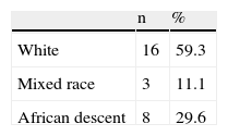
The first studies analyzing the types and upper limits of physiological adaptation to exercise were performed almost exclusively in white male athletes aged 18–35 years,13 these limits being universally accepted as reference criteria for differentiating such adaptations from pathological changes.
The limits have not been validated in populations of African or Afro-Caribbean descent, which may lead to incorrect diagnosis, with potentially serious clinical and personal consequences.
A study comparing adaptive limits between highly-trained athletes of African or Afro-Caribbean descent and whites14 showed that the mean increase in LV septal thickness was 12% greater in the former group, and that the prevalence of septal thickness >12 mm was 18% vs. 4%, with a maximum value of 16 mm in those of African descent and 14 mm in whites.
Although most of the changes in the heart induced by intense and prolonged sporting activity have now been thoroughly described, especially with regard to the left ventricle, variation over time of such changes requires clarification, particularly in terms of the response of the myocardium to seasonal variation in training intensity.
ObjectivesThe study had the following objectives: to assess variations in cardiac adaptation to exercise over a competitive season in individuals with previously induced adaptive changes, in order to determine whether additional changes occur in the echocardiographic parameters analyzed between the beginning of the training phase and the peak of the season; to determine whether there is a relationship between the magnitude of changes induced in particular cardiac structures and those in other structures; and to assess whether the adaptive pattern has similar characteristics in all athletes irrespective of ethnicity, age and percentage of participation in competitive games (an indicator of level of performance).
MethodsTwo assessments were performed by two-dimensional, M-mode and Doppler echocardiography in players in the senior squad of one of the top professional Portuguese football clubs at two different stages of the season:
- •
at the beginning of the training phase, following a period of partial deconditioning due to lack of supervised training and competitive games during the holiday period (a mean of 41.24 days);
- •
at the peak of the competitive season (February/March, the 7th and 8th months of the season), when the playing schedule is heaviest following the reconditioning characteristic of the early weeks of the season.
The choice of a professional team was dictated by the need to ensure intense daily training, supervised by staff who are experienced in training athletes for competition. It also ensures a high level of previous physical conditioning, particularly cardiovascular, induced by long periods of training and high-level competition.
All the athletes had previously undergone comprehensive evaluation by the club's medical department, and all were considered healthy.
Athletes who left the squad between the first and second assessments were excluded due to the lack of comparative data, as were those who joined between assessments.
All exams were performed by the authors, using the same equipment in all cases (Vivid i Cardiovascular Ultrasound System, General Electric® Healthcare, United Kingdom).
The exams were subsequently reviewed by a second observer, and in cases of disagreement, the parameters were jointly reviewed and discussed until a consensus was reached in order to eliminate the interobserver variability inherent to this imaging technique.
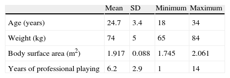
The players’ age, height, weight and body surface area (BSA) (using the Mosteller equation), ethnicity and number of years of professional playing were recorded at the beginning of the season. At the end of the season, the total time of participation in competitive games was calculated for each player, as well as that before and after the second echocardiographic assessment. Percentage of participation in competitive games is one aspect of an athlete's performance that can be quantified precisely.
The echocardiographic parameters assessed were LV wall thickness, LV mass (calculated using the Devereux formula), LVEDD, left atrial (LA) diameter, aortic root diameter, LA and right atrial (RA) areas, maximum right ventricular (RV) diameter, LV mass/BSA, E/A ratio, E/E′ gradient, tricuspid annular peak systolic velocity (TAPSV), ejection fraction (calculated by the modified Simpson method), RV-RA gradient, inferior vena cava diameter and respiratory variation, and estimated PASP.
The variables obtained in the first assessment were compared with those of the second for each player. The relationship between changes in echocardiographic parameters and demographic characteristics was then analyzed, as well as between the former and the percentage of participation in competitive games, with a view to determining any differences in the pattern of exercise-induced adaptations in the different subgroups.
Statistical analysisThe statistical analysis was performed using SPSS version 13.0 (SPSS Inc. Chicago, Ill).
Variables are expressed as means±standard deviation. The Student's t test, Wilcoxon test, Kruskal–Wallis test, and Spearman and Pearson correlation coefficients were used to compare variables and to infer statistical associations. A value of p<0.05 was considered statistically significant, with 95% confidence intervals.
A linear regression model, considering the percentage of participation in competitive games between the second echocardiogram and the end of the season as a dependent variable, and changes in septal thickness, LA diameter, RV-RA gradient and TAPSV as independent variables, was used to assess whether each independent variable was predictive of more competitive playing time during that period.
ResultsThe study population included 27 athletes. Demographic and anthropometric data and the results of the exams performed are shown in Tables 1–8.
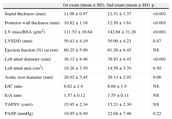
Changes in echocardiographic parameters.
| 1st exam (mean±SD) | 2nd exam (mean±SD) | p | |
| Septal thickness (mm) | 11.08±0.97 | 12.51±1.37 | <0.001 |
| Posterior wall thickness (mm) | 10.82±1.16 | 12.50±1.61 | <0.001 |
| LV mass/BSA (g/m2) | 111.52±16.64 | 142.68±31.26 | <0.001 |
| LVEDD (mm) | 50.43±4.19 | 50.90±4.23 | 0.47 |
| Ejection fraction (%) (at rest) | 60.25±5.99 | 61.26±6.45 | NS |
| Left atrial diameter (mm) | 36.12±4.46 | 38.93±4.43 | <0.001 |
| Left atrial area (cm2) | 19.26±3.59 | 19.56±3.70 | 0.50 |
| Aortic root diameter (mm) | 28.02±3.45 | 29.13±2.92 | 0.06 |
| E/E′ ratio | 6.82±1.4 | 6.68±1.4 | NS |
| E/A ratio | 1.57±0.12 | 1.55±0.11 | NS |
| TAPSV (cm/s) | 15.95±2.34 | 15.21±2.30 | NS |
| PASP (mmHg) | 19.95±6.49 | 22.68±7.48 | 0.22 |
BSA: body surface area; LV: left ventricular; LVEDD: left ventricular end-diastolic diameter; PASP: pulmonary artery systolic pressure; TAPSV: tricuspid annular peak systolic velocity.
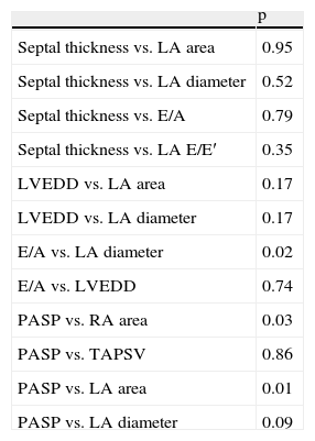
Statistical significance of correlations between changes in the parameters assessed.
| p | |
| Septal thickness vs. LA area | 0.95 |
| Septal thickness vs. LA diameter | 0.52 |
| Septal thickness vs. E/A | 0.79 |
| Septal thickness vs. LA E/E′ | 0.35 |
| LVEDD vs. LA area | 0.17 |
| LVEDD vs. LA diameter | 0.17 |
| E/A vs. LA diameter | 0.02 |
| E/A vs. LVEDD | 0.74 |
| PASP vs. RA area | 0.03 |
| PASP vs. TAPSV | 0.86 |
| PASP vs. LA area | 0.01 |
| PASP vs. LA diameter | 0.09 |
LA: left atrial; LVEDD: left ventricular end-diastolic diameter; PASP: pulmonary artery systolic pressure; RA: right atrial; TAPSV: tricuspid annular peak systolic velocity.
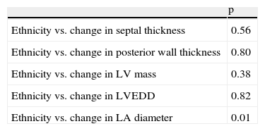
Differences in changes in parameters between athletes of different ethnicities.
| p | |
| Ethnicity vs. change in septal thickness | 0.56 |
| Ethnicity vs. change in posterior wall thickness | 0.80 |
| Ethnicity vs. change in LV mass | 0.38 |
| Ethnicity vs. change in LVEDD | 0.82 |
| Ethnicity vs. change in LA diameter | 0.01 |
LA: left atrial; LV: left ventricular; LVEDD: left ventricular end-diastolic diameter.
Correlation between changes in septal thickness and percentage of participation in competitive games.
| p | |
| Change in septal thickness vs. total % participation in competitive games | 0.04 |
| Change in septal thickness vs. % participation in competitive games before 2nd exam | 0.28 |
| Change in septal thickness vs. participation in competitive games between 2nd exam and end of season | 0.001 |
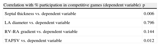
Analysis of possible predictors of percentage of participation in competitive games.
| Correlation with % participation in competitive games (dependent variable) | p |
| Septal thickness vs. dependent variable | 0.006 |
| LA diameter vs. dependent variable | 0.796 |
| RV-RA gradient vs. dependent variable | 0.144 |
| TAPSV vs. dependent variable | 0.012 |
LA: left atrial; RV-RA: right ventricular-right atrial; TAPSV: tricuspid annular peak systolic velocity.
Based on a regression model, considering the percentage of participation in competitive games between the second exam and the end of the season as a dependent variable, and changes in septal thickness, LA diameter, RV-RA gradient and TAPSV (selected using a stepwise method) as independent variables, the following results were obtained (Table 8):
DiscussionSignificant LV wall thickening was observed over the course of the playing season. At the peak of the season, 66.6% of athletes had a septal thickness of >12 mm, and 33.3% of >13 mm, with a maximum of 15.18 mm in a player of African descent. Differences in this parameter compared to other studies16 may be related to the particular sport studied and/or the fact that the second echocardiogram was performed at the peak of the season.
The fact that there was no statistically significant difference in LVEDD during the season suggests that a longer latency period is required for changes to be observed in this parameter on resting echocardiography.
Thus, in this type of population, subjected to chronic intense training that has resulted in profound adaptive changes, short-term seasonal variations in training intensity induce LV alterations, particularly in wall thickness and less so in chamber volume.
LV mass, which is derived from wall thickness and LVEDD, also increased between the first and second assessments. As expected, the same was observed with LV mass adjusted for BSA; at the first assessment, 63.0% of athletes had LV mass/BSA above the upper normal limit (102 g/m2),8 and 11.1% had criteria for severe LV hypertrophy. At the second assessment, only one player had LV mass/BSA below the upper normal limit and 55.5% had criteria for severe LV hypertrophy. Maximum LV mass was 279 g at the beginning of the season, and 458 g at the peak of the season.
Ejection fraction did not change significantly, since both assessments were performed under the same conditions of circulatory demand, and thus did not entail different conditions of preload and contractility.
Athlete's heart is characterized by normal diastolic function despite the presence of LV hypertrophy, which this study confirmed, since the hypertrophy observed between the two assessments did not impair diastolic function.
The LA, which acts as a conduit between the pulmonary veins and LV in diastole and as a reservoir in systole, has thinner walls than the LV and is thus more compliant and susceptible to chronic small changes in filling volume. There was a significant increase in LA diameters in the study population between the two assessments; however, the LA is not symmetrical15 and there is thus a difference in the magnitude of the increase in size depending on whether diameter or area is measured.
At the same time, there was a close correlation between increases in E/A ratio and LA diameter (Spearman coefficient 0.44, p=0.02), which suggests that the larger the LA, the greater the quantity of blood stored during ventricular systole, lengthening the E wave in the early diastolic phase of rapid LV filling. Since most ventricular filling occurs during this phase, the effect of greater atrial volume on the A wave is not as marked as on the E wave, resulting in a higher E/A ratio. In addition, as stated above, atrial contraction in trained athletes contributes less to ventricular filling than in sedentary controls, due to the former's lower heart rate and hence longer diastole.
On comparison of the two PASP measurements at rest in athletes with previous adaptive changes (under similar preload conditions), there was no statistically significant increase between the first and second assessments, despite a tendency for greater tricuspid regurgitation.
With regard to ethnic differences, players of African descent had greater LV wall thickness at the beginning and throughout the season, but this was not statistically significant due to the small sample.
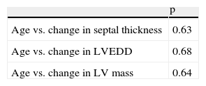
Age did not affect the patterns of additional adaptation, but once again this observation is of limited value due to the small number of athletes studied.
Thus, LV wall hypertrophy is the most important feature of additional adaptive changes between the beginning of the training phase and the peak of the season. It also appears to be related to performance, since in the linear regression model, increased septal thickness was an independent predictor of a higher percentage of participation in competitive games after the second assessment (p=0.008, by the F test).
ConclusionThe differences between the first and second assessments were slight, due to the fact that most of the morphological alterations found in athlete's heart had already occurred, the individuals studied having a long history of competition. The only differences found were in characteristics that tend to appear and disappear rapidly.
One important conclusion that can be drawn from the study is that additional adaptive changes take place over the course of the season depending on different levels of physical activity, mainly in LV wall hypertrophy, and consequent increased LV mass adjusted for BSA, and LA size. By contrast, variables such as LVEDD, ejection fraction and Doppler parameters of diastolic function do not change significantly.
The degree of septal thickening was shown to be an independent predictor of a higher percentage of participation in competitive games. This suggests that greater adaptive changes in the cardiovascular system are linked to better overall physical conditioning, reflected in the level of performance.
No particular characteristic, including hypertrophy and chamber dilatation, changed more in one group than in another.
To summarize, the characteristics of athlete's heart are not irreversible or static, but change according to the demands of the competitive season, and the magnitude of adaptive changes is not the same for all the parameters studied.
Conflicts of interestThe authors have no conflicts of interest to declare.
The authors are grateful to the Medical Department of Associação Académica de Coimbra-OAF, the cardiology department of Centro Hospitalar de Coimbra, and the National Cardiology Data Collection Center of the Portuguese Society of Cardiology.
Please cite this article as: Cabanelas N, et al. Evolução das características morfofuncionais do coração do atleta durante uma época desportiva. Rev Port Cardiol. 2013. http://dx.doi.org/10.1016/j.repc.2012.06.015.
