
Asymmetric basal septal hypertrophy is present in 10% of patients with hemodynamic significant aortic valve stenosis. From the surgeon's standpoint, it represents a dilemma as it may be implicated in suboptimal short and long-term results after aortic valve replacement (AVR), but also heighten unwarranted complications at the time of surgical correction. To provide insight about the usefulness and safety of concomitant septal myectomy in this setting, we performed a literature review searching Medline from its inception to November 2020 using the Pubmed interface. Only five low evidence retrospective analyses, comprising a total of <200 patients undergoing AVR with concomitant septal myectomy, were found in the literature. In summary, routine myectomy, in the presence of suspected or directly visualized asymmetric septal hypertrophy on echocardiogram during AVR, seems to be a safe procedure, with all authors reporting a low rate or absence of complications. Overall, myectomy in this setting is associated with superior echocardiographic results concerning surrogates of LV remodelling (LVM; LVM index; LVM/height) and diastolic function (E/E’), suggesting some benefit for hemodynamic outcomes. However, to what extent hemodynamic improvement is exclusively attributable to myectomy is uncertain, as is, the clinical significance of such an improvement, with similar short and mid-term survival rates being reported.
A hipertrofia assimétrica do septo basal está presente em cerca de 10% dos doentes com estenose aórtica hemodinamicamente significativa. Esta entidade representa um dilema do ponto de vista cirúrgico, uma vez que pode estar implicada em resultados subótimos após a substituição valvular aórtica, mas também em complicações, quando corrigida cirurgicamente. De forma a esclarecer a utilidade e segurança da miectomia quando realizada neste contexto, uma revisão da literatura foi realizada utilizando a Medline desde a conceção até novembro de 2020, através da interface Pubmed. Apenas cinco estudos retrospetivos de baixo nível de evidência foram encontrados. No seu conjunto totalizam menos de 200 doentes submetidos a substituição valvular aórtica e miectomia concomitante. Resumindo, a realização de miectomia por rotina na presença de hipertrofia septal assimétrica, identificada ecocardiograficamente ou por visão direta intraoperatoriamente, durante o procedimento de substituição valvular aórtica, é um procedimento aparentemente seguro, com todos os autores a reportarem taxas baixas ou ausentes de complicações relacionadas. De forma geral, a miectomia neste contexto foi associada a resultados ecocardiográficos, incluindo surrogates de remodelling ventricular esquerdo e função diastólica, superiores, sugerindo um benefício hemodinâmico. No entanto, qual o grau de melhoria que é atribuível isoladamente à miectomia é incerto, assim como é incerto o seu significado clínico, com sobrevida em curto e médio prazo equivalente reportada.
Left ventricular hypertrophy (LVH) in patients with aortic stenosis (AS) has long been recognized as an adaptive mechanism to long-standing pressure overload.1 Unlike primary causes, LVH in this setting is usually associated with uniform distribution. However, in a non-negligible number of patients undergoing aortic valve replacement (AVR), hypertrophy may present a non-uniform pattern, generally involving the basal portion of the septum.2 Asymmetric basal septal hypertrophy (ABSH) may contribute to residual left ventricular outflow tract (LVOT) obstruction which has been implicated in suboptimal short and long-term results after AVR.3–5
When faced with a patient with AS and ABSH, most surgeons perform only AVR. This decision is based on two assumptions: firstly, most surgeons believe that the normalization of LV afterload will in turn normalize LV hemodynamics and consequently promote LV remodeling; and secondly, the belief that avoiding an additional procedure might avoid potential related complications, such as atrioventricular block and septal perforation.6
However, a non-negligible number of surgeons perform additional concomitant septal myectomy (CSM), either liberally5,7 or in the presence of an subaortic obstruction identified on echocardiogram,2,8 in the presence of a predetermined interventricular septum (IVS) diameter cut-off value8; or based on in loco direct inspection of the LOVT.3,9
The rationale behind this more interventive approach is both the belief that CSM will improve long term results by optimizing afterload reduction, and that CSM may eliminate any residual dynamic subaortic gradient that can be unmasked by the correction of the valvular obstruction and heightened by post-operative hypovolemia and vasoactive drugs use.1,3,5
It is, therefore, important from the surgeon's standpoint, to clarify whether CMS results in: 1) superior long-term hemodynamic and clinical results; 2) better immediate post-operative course; or 3) no additional operative morbidity.
The aim of this paper is to review all the available evidence on the performance of concomitant myectomy for ABSH in patients undergoing AVR to provide surgeons with a clinical bottom line which they can use to guide their practice.
MethodsA comprehensive literature review was performed by searching Medline from its inception to November 2020 using the Pubmed interface. The following strategy was used: ((myectomy) OR (myotomy)) AND ((“aortic valve”) OR (“aortic stenosis”)). All 204 resulting articles were screened according to title and abstract. The references of relevant papers were screened for additional relevant publications. Articles pertaining to hypertrophic cardiomyopathy and congenital sub valvular obstruction were excluded. Five papers were found that provided evidence on the outcomes of myectomy in the specific setting of concomitant AVR.3,4,8–10 These results are presented in chronological order in the Results section and highlights summarized in Table 1.
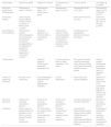
Highlights of the key characteristics and findings of the five papers relating to CSM in the setting of AVR.
| Author (date) | Tasca et al. (2003) | Kayalar et al. (2010) | Di Tommaso et al. (2013) | Lim et al. (2015) | Von Aspern et al. (2020) |
|---|---|---|---|---|---|
| Study type, patient group (CMS patients) | Retrospective study, n=196 (85) | Retrospective study, n=47 (47)No control group | Retrospective study, n=52 (29) | Retrospective study, n=301 (35) | Retrospective study, n=2199 (212)PSM (n=374) |
| Clinical key findings | Same actuarial survival (96.3%; CI 88.3%-99.3% vs. 98.8%; CI 96.4%-100%, p=0.806) | - | - | Same overall Survival (p=0.63) | Same mid-term survival (86.1±5% vs. 84.4±5%, p=0.957) |
| Key findings on echocardiogram | CMS presents superior echocardiographic results regarding left ventricular remodeling surrogatesCMS is an independent predictor of LVM/height regression at multivariate analysis | Significant reduction in LV dimensionsBut no control group present | CMS presents superior 5-years echocardiographic results regarding left ventricular remodeling and diastolic function surrogates | No difference in LV postoperative dimensions between the groups | - |
| Complications | - | Operative mortality: 2%Permanent pacemaker implantation:6% | No atrioventricular block or septal perforation in this cohort | Early death, AV block requiring pacemaker, stroke and renal failure, were statistically similar between the two groups | Same in-hospital mortality and pacemaker implantation rates. No iatrogenic VSD. |
| Criteria for performing myectomy | Not clear in the manuscript | Echocardiographic evaluation and surgical intraoperative inspection | Date of intervention | Direct visual and manual inspection | Based on HT discussion (IVS>14 mm; LVOT accelerated flow; risk of SAM)Final decision was at surgeon's discretion |
| Myectomy procedure | Similar to the technique described by Morrow11 | Similar to the technique described by Morrow11 | Similar to the technique described by Morrow11 | Not clear (described as standard myectomy) | Similar to the technique described by Morrow11 |
| Limitations | Small number of patients;Follow-up available in only 88% of surviving patients;Unclear clinical significance | No control group to compare results with AVR alone;Subjective criteria for performing myectomy. | Historical controls;Small number of patients;Unclear clinical significance | All procedures performed by the same surgeon;Small number of patients;Lack of regression measurements;Different group characteristics at baseline | Retrospective analysis;No clinical outcomes other than mortality;No hemodynamic outcomes. |
CSM: concomitant septal myectomy; CI: confidence interval; HT: heart team; IVS: interventricular septum; LVM: left ventricle mass; LVOT: left ventricle outflow tract; PSM: propensity score matched; SAM: systolic anterior motion; VSD: ventricular septal defect.
Tasca et al.4 observed 196 patients undergoing AVR, 85 of whom with CSM. Patients undergoing isolated AVR and AVR with CSM were retrospectively compared, and presented similar baseline characteristics (including type and size of implanted prosthesis and body surface area (BSA)), except for LVM/height ratio which was lower in the CSM group (78.6±19.8 g vs. 72.5±14.3 g; p=0.025). Actuarial survival at 2.9 years was similar between the two groups (96.3% vs. 98.8%; p=0.806) and no CSM-related complications were reported by the authors. In relation to the postoperative echocardiographic findings, the authors state that overall LV dimensions (including IVS thickness and mean LV thickness) decreased significantly in both groups, without any significant intergroup differences. However, LVM/height ratio (62.8±15.4 g vs. 55.7±12.8 g; p=0.002); LV thickness regression (-1.1±1.6 mm vs. -1.6±1.3 mm; p=0.016); and LVM regression after adjustment for covariates that correlated with mass regression (-16.8±17.8% vs. -24.6±14.7%; p=0.004), all decreased significantly more in the CSM group. Furthermore, multiple linear regression analysis showed that CSM was a powerful predictor of LVM/height regression (β=-0.306; p=0.001); with other predictors being body mass index and preoperative LVM/height ratio.
An interesting finding is that the CM group also presented significant lower mean transprosthetic gradient postoperatively (17.8±6.8 mmHg vs. 15.5±6.3 mmHg; p=0.034) which may present an additional confounding factor regarding LV remodeling. The clinical significance of these echocardiographic findings is, however, unclear, as no clinical endpoints are considered.
Kayalar et al.3 presented a retrospective cohort of 47 patients who underwent AVR and CSM at their institution between 2000 and 2009. The authors report statistically significant changes in LVM (211.4±54.3 g vs. 177.1±45 g; p<0.05); LVM index (113.7±24.3 g/m2 vs. 90.0±17.2 g/m2; p<0.05) and IVS thickness (13.9±2.5 mm vs. 11.4±1.6 mm; p<0.05), one year after combined AVR and CSM. Interestingly, the authors state that the potential need for myectomy was suspected in 28% of patients by preoperative echocardiography and in 28% of patients by intraoperative transesophageal echocardiography; but in the remaining 46%, the decision was based on direct inspection of the LVOT by the surgeon. The myectomy was performed in a similar fashion as for hypertrophic obstructive cardiomyopathy11 but only a mean of 0.8±0.61 g of muscle mass was excised. The authors report operative mortality of 2% and a 6% rate of permanent pacemaker implantation. An obvious limitation of this study is the lack of a comparison group. It is, therefore, impossible to assess the significance of the LV remodeling observed, for which AVR alone is, to a certain extent, responsible. Additionally, the indication for myectomy was highly subjective and surgeon-dependent, however, visual and digital inspection of the LVOT was the determining factor in the decision in this cohort.
Tommaso et al.9 reported the results of 52 patients with aortic stenosis and asymmetric septal hypertrophy (defined as LV septal/posterior wall thickness ratio of 1.3 or greater) retrospectively at their institution. All patients underwent AVR with a 21 mm mechanical prosthesis (St Jude or Carbomedics) and presented similar preoperative and operative characteristics, including similar five-year follow-up. The control group (AVR only) was operated on between 1997-1999 and the treatment group (AVR and CSM) was operated on between 2000-2004. The authors documented that LV remodeling (represented by left ventricular mass (LVM) regression, LVM index regression and intraventricular septum (IVS) thickness regression) and diastolic function (represented by E/E’ ratio regression) occurred in both treatment and control groups. However, both the absolute and percentual regression was superior in the treatment group: LVM regression (-9.3±3.3 g vs. -15±7.8 g; p=0.002); LVM index regression (-5.3±1.9 g/m2 vs. -8.8±4.2 g/m2; p=0.002); IVS regression (-1.5±0.9 mm vs. -2.0±0.8 mm; p=0.03); E/E’ ratio regression (-3.5±0.7 vs. -4.1±1.1; p=0.02). Furthermore, the authors report no atrioventricular block or septal perforation in their cohort, and conclude that the addition of myectomy to the standard AVR is safe in this setting.
When interpreting these results, one should bear in mind that historical cohorts may induce bias by disregarding time-related changes, other than the effective treatment itself. Additionally, the interpretation of the results should take into consideration the small number of patients involved. Although statistically significant, it remains to be seen whether the demonstrated improvement translates into any clinical significance.
Lim et al.8 retrospectively assessed 301 patients who underwent AVR and performed CSM (n=35) at the surgeon's discretion after intraoperative direct inspection of the LVOT.
The combined AVR and CSM group comprised a larger proportion of female patients (60% vs. 33.8%; p=0.0026); with smaller BSA (1.85±0.2 m2 vs. 1.97±0.2 m2; p=0.0049); higher left ventricular ejection fraction (LVEF) (63.2±13.3% vs. 55.4±15.2%; p<0.001); and smaller left ventricular end-systolic diameter (LVESD) (30.7±7.6mm* vs. 36.1±10.1mm*; p=0.0012) and left ventricular end-diastolic diameter (LVEDD) (48.8±6.0mm* vs. 52.1±8.3mm*; p=0.01). This group of patients was also target of a higher rate of implanted valves size ≤21 mm. Complications, including early and late death, AV block requiring pacemaker, stroke and renal failure, were statistically similar between the two groups. Postoperative echocardiographic measurements such as LV mass, LVM index, LVESD and LVEDD were not statistically different between the AVR alone and the CSM groups. Overall survival (OS) did also not differ between the two studied groups (p=0.63).
The authors sought to assess risk factors for CSM at the time of AVR. In the univariate analysis, they identified: the univariate analysis: gender (odds ratio (OR): 2.93; 95% confidence interval (CI): 1.42–6.04; p=0.004); BSA (OR:0.11; 95% CI:0.02–0.53; p=0.006); implanted valve size ≤21 mm (OR:3.2; 95% CI:1.54–6.65; p=0.002); LVEF (OR:1.05; 95% CI:1.01–1.08; p=0.008); LVESD (OR:0.93; 95% CI:0.88–0.98; p=0.005); LVEDD (OR:0.95; 95% CI:0.91–1; p=0.032); and LVOT/aortic-annulus ratio (OR:0.39; 95% CI:0.25–0.59; p<0.001) as potential predictors of CSM. At multivariate analysis (which excluded the LVOT/aortic-annulus ratio) only implanted valve size ≤21 mm (OR:3.2; 95% CI:1.54–6.65; p=0.002) was statistically significant. The authors highlight that the best cut-off point for preoperatively predicting CSM at the time of AVR is an LVOT/aortic-annulus <0.7.
Baseline differences between the two groups render direct comparison challenging and may underestimate the true effect of CSM on ventricular remodeling. The authors failed to document regression of LV dimensions which is arguably a more explicit surrogate of remodeling than final dimensions on their own. Additionally, the fact that all procedures were performed by the same surgeon makes generalization of the results more difficult.
Von Aspern et al.10 evaluated retrospectively 2199 patients undergoing either isolated AVR (n=1987) or AVR and CSM (n=212). Their analysis included a propensity matched subanalysis (n=374). No iatrogenic VSD was registered by the authors in this cohort. No difference was identified between the two groups regarding hospital mortality (2.1% vs. 1.6%, p=1.000), permanent pacemaker implantation rate (5.3% vs. 3.7%, p=0.62) and mid-term survival after 6 years (86.1±5% vs. 84.4±5%, p=0.957). Additionally, survival was similar to that expected for the overall population of the same region (p=0.178). In the multivariate analysis; patient age (hazard ratio (HR) 1.03; p=0.04) and small aortic annulus diameter (HR 1.42; p<0.001) were found to be independent predictors of CSM. Interestingly, the authors also present a survival analysis of patients that underwent isolated AVR stratified according to an end diastolic IVS cut-off value of 14 mm (isolated AVR IVS<14 mm, N=96 vs. IVS≥14 mm, N=91). The authors concluded that there was a trend toward inferior survival (78.1±8% vs. 89.7±5%; p=0.252) in patients with an IVS of ≥14 mm. As no clinical outcomes are presented other than OS, any midterm benefit from CSM is hard to establish. On the other hand, as no hemodynamic parameters were considered, any subclinical benefit of myectomy is also in doubt. Nevertheless, the safety of CSM was consistent with the previous publications.
ConclusionAsymmetric basal septal hypertrophy is found in about 10% of patients with hemodynamically significant aortic stenosis.2 The clinical significance of this finding is uncertain, which in turn leads to a surgical dilemma regarding how to approach it.
The answer to the question of whether routine concomitant myectomy provides superior outcomes in the setting of AVR is highly conditioned by the low quality of the available evidence. It comprises exclusively retrospective cohorts with heterogenous definitions, different study outcomes and varying follow-up periods. Furthermore, important factors contributing to LV remodeling, such as patient-prosthesis mismatch, size and type of prosthesis are not universally reported, making it impossible to attribute a causality to this procedure in this setting.
Due to a lack of prospective studies, no information is available on the impact of sub-aortic septal myectomy on the early post-operative course of AVR, namely the potential prevention of dynamic LVOT obstruction, in patients with low blood pressure receiving inotropes, in the setting of severe left ventricular hypertrophy.12,13
An additional unanswered question, not directly covered by the research strategy, is to identify patients at risk for persistent post-operative residual obstruction, after AVR, if sub-aortic myectomy is not performed and whether it contributes to incomplete functional improvement. Some authors have suggested cut-offs for LVOT area, sub-aortic interventricular septum thickness, narrowing of the distance between the septum and mitral valve in systole or reduced long LV axis/ascending aorta axis (sigmoid septum), systolic dagger-shaped Doppler spectrum, associated with an increased risk of residual subaortic obstruction.2
Considering the challenges related to transthoracic doppler differentiation between valvular and subvalvular obstacles, cardiac magnetic resonance may emerge as the modality of choice to identify patients at risk.14
With that said, routine myectomy, in the presence of asymmetric septal hypertrophy suspected or directly visualized on echocardiogram during AVR, seems to be a safe procedure, with all authors reporting a low or absent complications rate. Moreover, myectomy in this setting is associated with superior echocardiographic results concerning surrogates of LV remodeling (LVM; LVM index; LVM/height) and diastolic function (E/E’), suggesting some benefit for hemodynamic outcomes. However, the extent to which hemodynamic improvement is exclusively attributable to myectomy is uncertain, as is the clinical significance of such an improvement. A large prospective randomized comparison study is warranted to provide further evidence so cardiac surgeons can follow the best conduct in the setting of ABSH and AVR.
P.L. Magro and M. Sousa Uva's personal opinion, based on their own subjective experience, is that liberal CSM, especially in patients with high-risk characteristics for dynamic obstruction (sigmoid septum; basal septum width>15 mm; visually identifiable basal bulging) provides a potential hemodynamic advantage in the post-operative period and may facilitate long-term LV remodeling.
Conflicts of interestThe authors have no conflicts of interest to declare.
