
The number and complexity of cardiac implantable electronic devices (CIEDs) have increased, as has the number of related complications, often leading to removal of the implanted system. The aim of this study was to characterize transvenous explantation/extraction of CIED leads in a reference center.
MethodsThis was a descriptive observational study of patients consecutively admitted from January 2009 to May 2014 for transvenous lead extraction.
ResultsThe sample consisted of 109 patients, with a mean age of 64.6±16.62 years, 73.1% male. The main indication for lead extraction was CIED infection. The mean time from first implantation to lead removal was 5.6±4.89 years. Blood cultures were positive in 32.8% of cases and 29% of patients had vegetations on echocardiography. A total of 228 cardiac leads were removed, of which 58.8% were ventricular, 32.4% atrial and 8.8% coronary sinus. Complete clinical success was achieved in 97.2% of cases, while procedural success was complete in 93.4% and partial in 5.3%. The complications reported were three cases of significant pocket hematoma, one of subclavian vein thrombosis and three of cardiac tamponade, effectively treated by pericardiocentesis.
ConclusionsTransvenous explantion or extraction of CIED leads was highly effective. A high level of experience is an essential factor in the success and safety of the procedure.
O número e a complexidade dos dispositivos cardíacos implantáveis têm vindo a aumentar assim como as suas complicações, conduzindo frequentemente à remoção dos mesmos. O objetivo deste estudo foi caraterizar a população envolvida e os procedimentos utilizados na remoção transvenosa de eletrocateteres cardíacos num centro de referência.
MétodosEstudo observacional descritivo dos procedimentos de remoção transvenosa de eletrocateteres cardíacos de janeiro de 2009 a maio de 2014.
ResultadosA amostra incluiu 109 doentes, com idade média de 64,6 anos (± 16,62), 73,1% do género masculino. A principal indicação para remoção de eletrocateteres foi infeção do dispositivo. O tempo médio desde a primeira implantação à remoção foi de 5,6 anos (± 4,89). As hemoculturas foram positivas em 32,8% dos casos e cerca de 29% dos doentes apresentavam no estudo ecocardiográfico vegetações. Foram removidos 228 eletrocateteres, 58,8% ventriculares, 32,4% auriculares e 8,8% do seio coronário. O sucesso clínico do procedimento foi completo em 97,2%; o sucesso do procedimento foi completo em 93,4% dos casos e parcial em 5,3%. Como complicações registaram-se três hematomas volumosos da loca, uma trombose da veia subclávia e três tamponamentos cardíacos, tratados eficazmente com pericardiocentese.
ConclusõesA explantação/extração transvenosa dos eletrocateteres mostrou-se altamente eficaz. Contudo, experiência com o sistema é um fator preponderante para o sucesso e segurança do procedimento.
The number and complexity of cardiac implantable electronic devices have increased, leading to a growing incidence of related complications.1 There has thus been a significant increase in procedures to remove pacemakers, implantable cardioverter-defibrillators (ICDs) and coronary sinus leads, the most common reason being device infection.2–4 Various techniques are used for device extraction, all associated with a significant risk of complications.The aim of this study was to characterize device removal procedures in a reference center.
MethodsThis was a descriptive observational study of patients consecutively admitted to a reference center for lead extraction from January 2009 to May 2014.
Pocket infection was defined as the presence of an abscess, purulent drainage, suture dehiscence, skin erosion or skin adherence. Device-related endocarditis was defined as persistent bacteremia or sepsis in the absence of another identifiable source or vegetations on the device.All patients undergoing lead extraction were informed of possible complications and gave written informed consent.
The procedures were performed in the pacing and electrophysiology unit of the cardiology department, with cardiothoracic surgical backup in case a patient required emergent surgical intervention, under sedoanalgesia and continuous hemodynamic and electrocardiographic monitoring.
Cook Medical devices were used for the extraction procedures, this choice being dictated by their availability at the center and by the medical team's experience with these techniques of lead extraction. In all cases, an initial attempt was made to remove the leads by simple traction; if this was unsuccessful, a Liberator® Beacon® Tip Locking Stylet (Cook Medical Inc., Bloomington, USA) was employed, followed by an Evolution® RL Controlled-rotation Sheath Dilator Set (Cook Medical Inc., Bloomington, USA), which has a manually rotated steel tip for tissue debridement.Patients remained in the cardiac intensive care unit for 24 hours after the procedure.
Procedural and clinical success were defined according to the 2009 Heart Rhythm Society (HRS) Expert Consensus on transvenous lead extraction.3 Complete procedural success was defined as removal of all targeted leads and all lead material from the vascular space, with the absence of any permanently disabling complication or procedure-related death. Retention of a small part (≤4 cm) of the lead (coil or insulation) of the lead was considered partial success. Clinical success was defined as removal of all targeted leads and all lead material from the vascular space, or retention of only residual material that does not negatively impact the outcome goals of the procedure. Failure was defined as inability to achieve either complete procedural or clinical success, or the development of any permanently disabling complication or procedure-related death. Major and minor complications were defined according to the HRS expert consensus statement.3
Patient and procedural data were collected, including demographic characteristics, echocardiographic data, laboratory and blood culture results, lead removal technique and complications. The statistical analysis was performed using SPSS version 17.0.
ResultsThe study included 109 patients, with a mean age of 64.6±16.62 years, 73.1% male. In one case the reason for extraction was lead fracture, in another the patient requested removal of an ICD implanted for primary prevention for psychological reasons, while in the remaining cases the reason was device infection. The mean time from first implantation to lead extraction was 5.6±4.89 years (3 weeks–24 years).
Type 2 diabetes was present in 26.8% of the sample; 25% had been under anticoagulant therapy and 5.5% had had significant pocket hematoma. Since a significant proportion of the patients had been transferred from other hospitals, several had undergone bridging with low molecular weight heparin, while others were under warfarin at the time of the procedure. Signs of pocket infection (erosion, adhesion, erythema, purulent drainage, device or lead exteriorization) were seen in 63.8% of cases, and three patients presented in septic shock. Around 36% of patients had a history of previous infection treated conservatively (with antibiotic therapy or generator replacement, or by removing the generator, cutting the leads, and reimplanting on the contralateral side). Laboratory tests showed elevated C-reactive protein (CRP) (>5.0 mg/l) in 63% of patients, although with only slight rises in most cases (mean 4.8 mg/l). Leukocytosis was seen in 11.7%, renal failure in 31.6% and hemoglobin below reference values in 30%. Blood cultures were positive in 32.8% of cases, and vegetations were observed on transthoracic and/or transesophageal echocardiography in 29%, the largest of which was 30 mm at its widest point. Echocardiographic assessment revealed 52% of patients had impaired left ventricular systolic function.
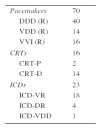
The procedures in the 109 patients involved the removal of 228 leads: 58.8% right ventricular, 32.4% atrial and 8.8% coronary sinus. The devices removed were 70 pacemakers, 16 cardiac resynchronization devices (two cardiac resynchronization therapy pacemakers [CRT-P] and 14 cardiac resynchronization therapy defibrillators [CRT-D]) and 23 ICDs (Table 1). Extraction of leads was performed in 84.3% of cases and simple explantation in the others. The mean times from implantation to explantation and to extraction were 0.39±0.28 years and 6.78±4.7 years, respectively. In patients who had been implanted less than one year previously (2.8%), 62% of procedures were explantations. One extraction of a fractured lead was carried out by femoral access using a Needle's Eye Snare® (Cook Medical Inc., Bloomington, USA), by pulling the lead after its free end had been snared. All the other leads were removed by the same superior venous access used for implantation: 17.5% of leads were removed by simple traction after inserting a conventional guidewire, 6.5% with the Liberator® Beacon® Tip Locking Stylet (Cook Medical Inc., Bloomington, USA), and the others (the majority) using the Evolution® sheath (Cook Medical Inc., Bloomington, USA).
Cardiac implantable electronic devices removed in the study population.
| Pacemakers | 70 |
| DDD (R) | 40 |
| VDD (R) | 14 |
| VVI (R) | 16 |
| CRTs | 16 |
| CRT-P | 2 |
| CRT-D | 14 |
| ICDs | 23 |
| ICD-VR | 18 |
| ICD-DR | 4 |
| ICD-VDD | 1 |
CRT: cardiac resynchronization therapy device; CRT-D: cardiac resynchronization therapy with defibrillator; CRT-P: cardiac resynchronization therapy with pacemaker; ICD: implantable cardioverter-defibrillator.
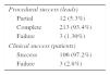
Clinical success was achieved in 97.2% of patients, complete procedural success in 93.5% and partial success in 5.3%. The procedure failed in only three cases, in which right ventricular leads could not be extracted (Table 2).
There were few complications: three cases of cardiac tamponade, effectively treated by pericardiocentesis; one small pericardial effusion without hemodynamic compromise; one case of subclavian vein thrombosis; and three of significant pocket hematoma.
DiscussionDevice infection is the leading indication for removal of cardiac implantable electronic devices.3,5 The increasing number and complexity of these devices, together with an aging target population with more comorbidities, are leading to a growing incidence of infections.1 Greenspon et al. conducted a descriptive analysis of the infection burden for pacemakers and ICDs in the USA and found that over a 16-year period the incidence of four major comorbidities (renal failure, heart failure, respiratory failure, and diabetes) increased in parallel with increases in numbers of device implantations (96%), in infection rate (from 1.53% to 2.41%) and in mortality associated with device infection (from 2.91% to 4.69%). Risk factors for infection included reintervention to replace a depleted battery and implantation of cardiac resynchronization systems or ICDs, due to their longer life and hence need for more generator replacements.1 Similarly, in our population there were high prevalences of type 2 diabetes (26.8%), renal failure (31.6%) and impaired left ventricular systolic function (51.5%). In a follow-up of patients four years after first CRT-D implantation in an Italian registry, Landolina et al. reported that 50% of patients required surgical revision for battery depletion and 14% for unanticipated events. The infection rate was 1%/year, and the risk of infection increased after device replacement procedures.6 It is thus to be expected that with widening indications for implantation of cardiac resynchronization systems, the incidence of complications – and therefore of extractions – will increase.
Infection of cardiac electronic devices has a wide spectrum of manifestations, ranging from pocket infection to sepsis and death. Pocket infection may present with skin erosion or erythema, abscess, purulent drainage and/or suture dehiscence, and may be accompanied by local pain with no other initial symptoms.1,3–5 In our study 63.8% of patients presented signs of pocket infection; previous infection was identified in 36%, who had been treated conservatively on the basis of the patient's general condition and comorbidities. As the study was conducted in a reference center for extraction of cardiac electronic devices, many patients had been referred from other centers and no information was available on the reasons for their initial conservative treatment. The 2000 North American Society of Pacing and Electrophysiology guidance document stated that it was acceptable to remove the device and cut the exposed parts of the leads in cases of pocket infection in which the intravascular portion of the system was not involved. However, such a strategy is proving to be unsuccessful, with a high risk of recurrence of reinfection and potentially serious consequences such as device-related or valvular endocarditis and sepsis, putting the patient at unacceptable risk. Mortality in cases of persistent infection can be as high as 66% if the leads are not removed.7 The HRS consensus document recommends complete removal of the system (device, leads, adapters, sutures) and as much of the infected tissue as possible when an infection is identified, even if only of the pocket. However, in specific situations, particularly when the patient has a poor prognosis or comorbidities that carry an excessively high risk of complications, it is acceptable to adopt a more conservative strategy.2
Although most device infections involve only the pocket, around 10% manifest as endocarditis.8 In our study, blood cultures were positive in 32.8% of cases and vegetations were identified by transthoracic and/or transesophageal echocardiography in 29% of patients. According to the literature, inflammatory markers are not reliable indicators of infection, since they may be normal even in the presence of endocarditis.1,5 Laboratory tests in our population showed elevated CRP in 63% of patients, although with only slight rises in most cases (mean 4.8 mg/l); leukocytosis was seen in 11.7%. One reason that these patients often did not present high levels of inflammatory markers or positive blood cultures is that they had been taking antibiotics prior to the diagnosis of device infection.
Studies have identified various risk factors for device infection, including diabetes, heart failure, renal failure, oral anticoagulation, chronic corticosteroid therapy, fever 24 hours before device implantation, temporary transvenous pacing prior to implantation of a permanent pacemaker, early reintervention, failure to use prophylactic antibiotic therapy before the procedure, operator experience, device revision or replacement, and amount of indwelling hardware. In a Danish study of 46299 patients, Johansen et al. showed that repeat procedures were associated with a substantial rise in risk of infection; independent predictors of pacemaker infection were the number of interventions (including replacements), male gender, younger age, implantation during the early stages of the study, and absence of antibiotic therapy, while female gender, older age, and antibiotic prophylaxis before the procedure were associated with lower risk of infection.9
Lead extraction is a technically challenging procedure. After initial implantation, areas of thrombus form along the leads that then organize and fibrose, hampering extraction. In general, the longer the lead has been in place, the greater the extent of fibrosis, further complicating removal. Lead removal involves not only retracting the lead from its attachment to the myocardium, but also freeing the lead from the fibrous sheath that anchors it to the vasculature.9,10 The points most likely to fibrose include the initial entry to the subclavian vein, the superior vena cava and the lead/myocardial interface, although there are often several areas of adherence along the vein that hinder extraction. Calcification of the fibrous tissue may occur over time, especially in young patients.10 The presence of multiple leads and the time since implantation are critical factors in the success of the removal procedure. In our study, simple explantation was associated with shorter mean time since implantation (0.39±0.28 years) than extraction (6.78±4.7 years). In the subgroup with less than one year since implantation, the device was removed by simple traction in 62% of cases.
There are various extraction techniques, all associated with potentially serious complications, including death. Early techniques involved simple manual traction that frequently proved ineffective and carried a high risk of complications including myocardial avulsion, tamponade, and death. The past 30 years have witnessed significant advances in lead extraction technology, resulting in safer and more efficacious techniques.10 A 2013 study on 206 lead extraction procedures in 122 patients showed comparable procedural success and safety in transvenous laser extraction and a mechanical approach, and clinical success and cost-effectiveness analysis favored the mechanical approach.11 In the majority of cases leads are extracted by the same venous access as used in implantation (superior approach) by simple traction or using locking stylets and special sheaths. If the leads are not accessible via the implant site, particularly if they had been previously cut and retracted into the venous system, femoral or jugular access are options.12–14 Bongiorni et al. described a method used to extract 2062 leads with a single mechanical dilating sheath via a subclavian approach but using a femoral approach combined with an internal transjugular approach when the superior approach failed, with a high success rate and few complications.15 In our population, only one extraction was by the femoral approach, using a Needle's Eye Snare® (Cook Medical Inc., Bloomington, USA); in all other cases the superior approach was used.
Nowadays the success rate for transvenous extraction is high, with few complications.11,16 Nevertheless, complications can be serious or even fatal. Whatever technique is used, a high level of operator experience is essential for the success of the procedure. In our study there were only three major complications, pericardial effusion with tamponade, effectively treated by pericardiocentesis; emergency surgical intervention was not required in any of the cases. Even so, given the possible need for emergent intervention, cardiothoracic surgical backup should be available at all times.
ConclusionsThere has been an exponential increase in the number and complexity of cardiac implantable electronic devices. The need for removal of these devices is also increasing, by explantation or extraction, in most cases due to device infection. All available techniques are associated with significant risk of complications, most of them minor, but potentially fatal complications can arise. In our experience the removal techniques used were effective, with few complications.
Ethical disclosuresProtection of human and animal subjectsThe authors declare that no experiments were performed on humans or animals for this investigation.
Confidentiality of dataThe authors declare that no patient data appears in this article.
Right to privacy and informed consentThe authors declare that no patient data appears in this article.
Conflicts of interestThe authors have no conflicts of interest to declare.
Please cite this article as: Ribeiro S, Leite L, Oliveira J, Pereira MJ, Pinheiro C, Ermida P, et al. Extração transvenosa de eletrocateteres de dispositivos cardíacos implantáveis. Rev Port Cardiol. 2015;34:739–744.
