Informação da revista
Vol. 33. Núm. 5.
Páginas 317-318 (Maio 2014)
Partilhar
Partilhar
Baixar PDF
Mais opções do artigo
Vol. 33. Núm. 5.
Páginas 317-318 (Maio 2014)
Image in cardiology
Open Access
Mediastinal hydatid cyst compressing the pulmonary artery and atrial septal defect: A rare association
Cisto hidático do mediastino comprimindo a artéria pulmonar e defeito septal auricular – uma associação rara
Visitas
5130
Arif Sunera, Sedat Koroglub,
Autor para correspondência
k.sedat@mynet.com

Corresponding author.
, Musa Cakicia, Mustafa Polata, Mustafa Cetina, Bulent Petikc, Recai Kayad, Hakan Kayaa
a Adiyaman University, Education and Research Hospital, Cardiology Clinic, Adiyaman, Turkey
b Afsin State Hospital, Cardiology Clinic, Kahramanmaras, Turkey
c Adiyaman University, Education and Research Hospital, Radiology Clinic, Adiyaman, Turkey
d Adiyaman University, Education and Research Hospital, Anesthesiology Clinic, Adiyaman, Turkey
Este item recebeu

Under a Creative Commons license
Informação do artigo
Texto Completo
Bibliografia
Baixar PDF
Estatísticas
Figuras (3)
Mostrar maisMostrar menos
Texto Completo

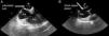
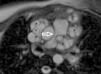
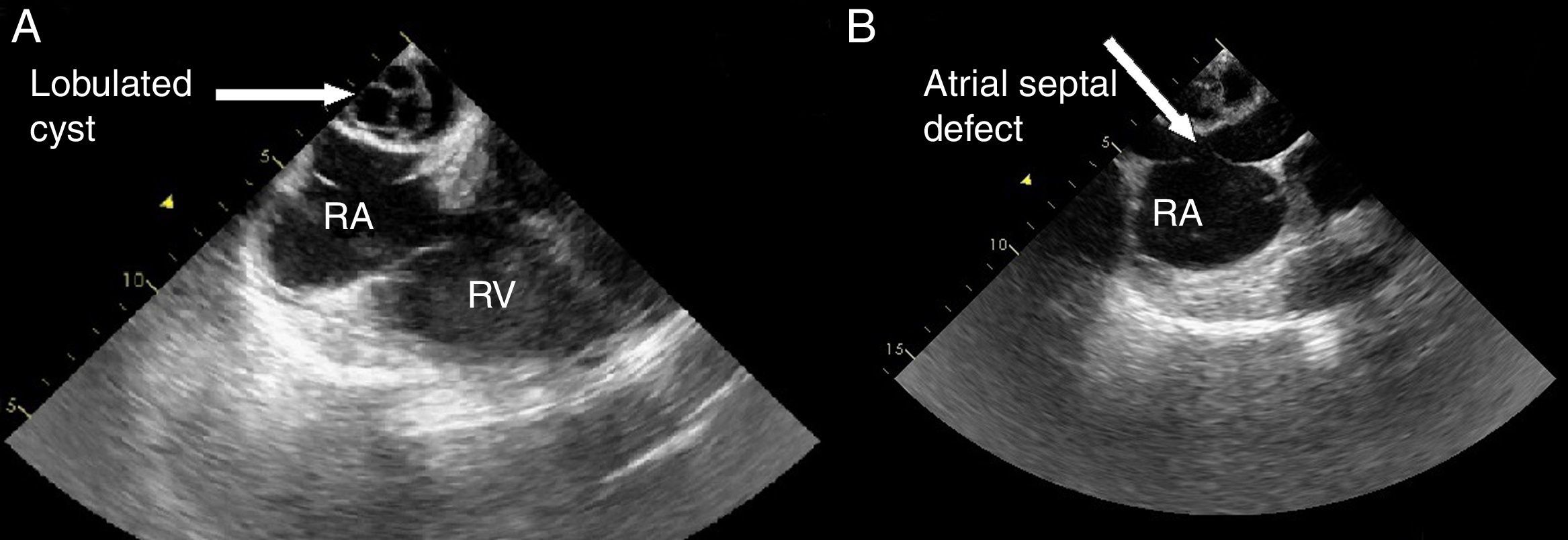
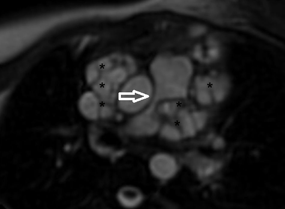
A 21-year-old woman was admitted to our clinic with dyspnea. Her medical history was unremarkable. Right bundle branch block was identified on 12-lead ECG and mediastinal enlargement on the chest X-ray. Transthoracic and transesophageal echocardiographic examination showed a lobulated cystic image in the left atrium and secundum-type atrial septal defect (Figures 1 and 2). Additionally, in short-axis view a mass was visualized invading the pulmonary artery, and continuous wave Doppler of the pulmonary artery revealed a maximum gradient of 40 mmHg. A detailed examination with cardiac magnetic resonance imaging demonstrated a multicystic mass in the mediastinum compressing the main pulmonary artery, predominantly the left branch (Figure 3). The patient was diagnosed with secundum atrial septal defect and mediastinal hydatid cyst on the basis of serologic tests and detailed history. Surgical therapy was offered and she asked for time to think. Medication with albendazole was started, but she was then lost to follow-up.

Figure 1.

Transesophageal echocardiographic views showing (A) lobulated cyst and (B) atrial septal defect. RA: right atrium; RV: right ventricle.

(0,15MB).
Figure 2.

Transthoracic echocardiographic views showing (A) multiple cystic mass compressing the pulmonary artery and (B) continuous wave Doppler of the pulmonary artery with maximum gradient of 40 mmHg. PA: pulmonary artery.

(0,11MB).
Figure 3.

Cardiac magnetic resonance imaging showing external compression of pulmonary artery. Black asterisks indicate hydatid cysts.

(0,05MB).

Hydatid cyst is a parasitic disease caused by Echinococcus granulosus. The predominantly affected organs are the liver and the lung; mediastinal location is very rare.1 The preferred method for management is surgery but adjunctive albendazole therapy is the subject of debate.

Ethical disclosuresProtection of human and animal subjects

The authors declare that no experiments were performed on humans or animals for this study.

Confidentiality of data

The authors declare that they have followed the protocols of their work center on the publication of patient data.

Right to privacy and informed consent

The authors have obtained the written informed consent of the patients or subjects mentioned in the article. The corresponding author is in possession of this document.

Conflicts of interest

The authors have no conflicts of interest to declare.

Reference
[1]
T. Isitmangil, A. Toker, S. Sebit, et al.
A novel terminology and dissemination theory for a subgroup of intrathoracic extrapulmonary hydatid cysts.
Med Hypotheses, 61 (2003), pp. 68-71
Copyright © 2013. Sociedade Portuguesa de Cardiologia
Idiomas
Revista Portuguesa de Cardiologia
Opções de artigo
Ferramentas
en pt

Are you a health professional able to prescribe or dispense drugs?

Você é um profissional de saúde habilitado a prescrever ou dispensar medicamentos

Ao assinalar que é «Profissional de Saúde», declara conhecer e aceitar que a responsável pelo tratamento dos dados pessoais dos utilizadores da página de internet da Revista Portuguesa de Cardiologia (RPC), é esta entidade, com sede no Campo Grande, n.º 28, 13.º, 1700-093 Lisboa, com os telefones 217 970 685 e 217 817 630, fax 217 931 095 e com o endereço de correio eletrónico revista@spc.pt. Declaro para todos os fins, que assumo inteira responsabilidade pela veracidade e exatidão da afirmação aqui fornecida.