
Infective endocarditis is a common complication among injecting drug users. Disease risk among these patients is increased by the spread of HIV infection. In the following article, we discuss the exceptional clinical presentation of a 28-year-old patient who used intravenous drugs (heroin) for 10 years, had been infected with HIV for seven years and as a complication had developed Staphylococcus aureus infective endocarditis.
The patient came to the hospital in serious condition, complaining of bodily pain, swelling of the legs and general weakness. During hospitalization, besides infective endocarditis, she was also diagnosed with anemia, toxic hepatitis, renal failure, ascites, sepsis, and pneumonia. A completely disrupted tricuspid valve, damaged aortic valve, and fibrosis of the mitral valve were detected. Echocardiographic and radiologic data showed that the patient's condition continued to deteriorate day by day, with significant progression of heart failure, ejection fraction decreasing from 45% to 10%, and development of myocarditis, hydrothorax and pericarditis. However, this progressive worsening of the patient's condition ceased when vancomycin was administered. To the authors’ knowledge, this is the first such case described in the literature in which significant improvement was observed despite the patient's complex condition with associated complications.
A endocardite infecciosa é uma complicação comum entre os consumidores de drogas injetáveis. O risco desta doença nestes pacientes aumenta com a propagação da infeção do VIH. Neste artigo, discute-se a apresentação clínica de uma jovem doente que consumiu drogas intravenosas (heroína) durante dez anos e que há sete anos que está infetada pelo VIH. Para além disso, desenvolveu endocardite infecciosa causada por Staphylococcus aureus. De 28 anos de idade apresentou-se no hospital em estado grave, queixando-se de dores no corpo, pernas inchadas e de um estado geral de fraqueza.
Durante o internamento, para além da endocardite infecciosa, foi-lhe também diagnosticado – anemia, hepatite tóxica, insuficiência renal, ascite, sepsis e pneumonia. Foram também detetadas interrupção total da válvula tricúspide, válvula aórtica danificada e fibrose da válvula mitral. De acordo com a avaliação ecocardiográfica e dados radiológicos, a situação da doente continuou a deteriorar-se: a insuficiência cardíaca progrediu significativamente, a fração de ejeção diminuiu de 45% para 10% e desenvolveram-se miocardite, hidrotórax e pericardite. No entanto, apesar da situação grave da doente progredir de modo contínuo, houve uma melhoria assim que lhe foi administrada vancomicina. De acordo com os autores, este será o primeiro caso clínico descrito na literatura que revelou uma melhoria significativa apesar da situação complexa da doente com demais complicações.
aortic valve
congestive heart failure
C-reactive protein
ejection fraction
erythrocyte sedimentation rate
heart failure
intensive care unit
intravenous drug users
infective endocarditis
left ventricle/left ventricular
methicillin-resistant Staphylococcus aureus
mitral valve
red blood cells
right coronary
right ventricle/right ventricular
tricuspid valve
Despite advances in diagnosis, infective endocarditis (IE) remains a common cause of hospitalization, with high morbidity and mortality rates.1 This may be due to the changing epidemiology of IE, including increasing antimicrobial resistance, more frequent heart surgery and prosthetic valve implantation, and widespread use of intravenous drugs.2
IE was initially recognized to be a complication of intravenous drug use in the 1950s.3 It is a potentially fatal consequence of injecting illicit drugs, such as heroin, cocaine, and methamphetamine. Intravenous drug use increases IE risk through a variety of mechanisms. Drug solutions may contain particulate matter (e.g. talc) that damage cardiac valves if injected intravenously. In addition, poor injection hygiene and injecting contaminated drug solutions can introduce high circulating bacterial loads.4 In intravenous drug users (IDUs), the incidence of IE is 2%–5% and it is responsible for 5%–10% of deaths.5
HIV infection increases the risk of IE. In developed countries HIV infection among IDUs with IE ranges between 30% and 70%.6 The tricuspid valve (TV) may be more susceptible to heroin use, as heroin can cause an increase in pulmonary arterial pressure, creating more turbulence at the TV.7
In this article we describe an exceptional clinical case of an IE patient who was HIV-infected and also injected heroin. We compare the case with data in the literature and analyze the differences, and also review the clinical features of IE among IDUs, nonaddicts, and HIV-infected and uninfected patients.
Case reportA 28-year-old Lithuanian woman was admitted to our hospital. Her presenting complaints were bodily pain and general weakness. She had been addicted to heroin for 10 years. Her past medical history was significant for anemia and chronic pyelonephritis, leading to stage 2 renal failure, and infection with HIV for seven years. She was an alcohol abuser and a smoker. Physical examination on presentation showed body temperature of 36.8°C, edema in the legs, pale mucous membranes, dry tongue, normal cardiac rhythm and breath sounds, respiratory rate 20/min, heart rate 88 beats/min, and blood pressure 120/80 mmHg. Laboratory tests revealed microscopic hematuria and leukocyturia and C-reactive protein (CRP) 28 mg/l, erythrocyte sedimentation rate (ESR) 45 mm/h, and hemoglobin 59 g/l. Ascites was found in the abdomen. X-ray study showed no abnormalities in the thoracic organs. On day 7 of hospital stay the patient's condition deteriorated. The X-ray showed moderate bilateral hydrothorax, with a slightly sharpened image of the lungs and an interstitial component, local infiltration of the right cardiodiaphragmatic angle, inflammatory lesions around the pleura on the left, and enlarged cardiac diameter in both directions. On day 8 echocardiography was performed, which detected vegetations on the TV and the right atrium. C-reactive protein was elevated (38.00 mg/l). Before the administration of antibiotics Staphylococcus aureus (sensitive to penicillin, oxacillin and erythromycin) was detected in blood culture and Escherichia coli (sensitive to ciprofloxacin and gentamicin) in urine culture. On the following day the patient was given ampicillin and ceftriaxone (Table 2), by which time her temperature had risen to 38.8°C. After a few days echocardiography was repeated in order to assess changes, and showed decreasing ejection fraction (EF) and increasing pericardial effusion (Table 1, 11 days of treatment). Antibiotic therapy was changed to ceftriaxone (Oframax) only (Table 2). Sepsis, pneumonia, toxic hepatitis and infectious endocarditis were additionally diagnosed. The blood analysis was repeated, showing CRP 30.00 mg/l and white blood cell count 7.4×10⁹/l. The patient's condition continued to deteriorate, with breathlessness and anasarca; an audible grade 3 systolic murmur along the parasternal line and mild congestive rales in both lower lung fields were detected. Secondary myocarditis was diagnosed. Serum CD4+ cell count was 651/mm3, and antiviral therapy for HIV was not initiated. In order to assess the effect of antibiotic therapy, blood and urine cultures were re-evaluated. On day 8 bacteria were not detected in urine culture, but S. epidermidis was detected in blood cultures. Antibiotic therapy was changed to a combination of oxacillin and gentamicin (Table 2). On day 20 the echocardiogram showed significantly decreasing EF (10%) (Table 1). The ECG showed sinus rhythm and inverted T waves in leads I, aVL, V5, and V6. An elevated D-dimer level (2.25 μg/ml) was detected. Repeat chest X-ray showed progression of pleuritic lesions on the right (hydrothorax), the right lung being almost completely covered with liquid, and the mediastinum displaced to the left, covering the lower left lung field. While still in a critical condition, the patient was given vancomycin (Table 2), which led to regression of the abnormalities, bacteria being undetectable in blood culture. On day 44 the ECG showed sinus rhythm, and there was reversal of T-wave inversion in leads I, aVL, and V5-V6. On the chest X-ray the hydrothorax had disappeared. After treatment with vancomycin, echocardiography revealed a significant recovery of EF (from 10% to 50%) (Table 1, 48 days of treatment).
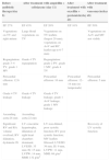
Evolution of echocardiographic changes according to antibiotic therapy.
| Before antibiotic therapy(day 8) | After treatment with ampicillin + ceftriaxone (day 11) | After treatment with oxacillin + gentamicin(day 20) | After treatment with vancomycin(day 48) | |
|---|---|---|---|---|
| EF 27% | EF 45% | EF 20% | EF 10% | EF 50% |
| Vegetations on TV and right atrium | Large florid vegetations on TV | Vegetations on TV leaflets (largest 29 mm), vegetations on AoV and RC leaflet (up to 6-7 mm) | Vegetations on AoV and MV not visible | |
| Regurgitation: grade 3–4 TV, grade 1 MV | Grade 4 TV regurgitation | Regurgitation: grade 2 PV, grade 1 MV, grade 4 TV | ||
| Pericardial effusion: 12.6 mm | Pericardial effusion: 16 mm | Pericardial effusion: 12–14 mm (no tamponade) | Pericardial effusion: 5 mm | |
| Grade 4 TV leakage | Grade 4 TV leakage | Grade 4 TV leakage, grade 1 AoV leakage, grade 1 MV leakage | ||
| Ascending aorta:28 mm | Ascending aorta:22 mm | |||
| Significant RV and RA volume overload, RV dilatation, dyskinetic IS | LV concentric hypertrophy, dilatation of right heart chambers dominant; LVESD: 35 mm; IS: 15 mm; LVPW: 14 mm; MMI: 132 g/m2 | LV non-dilated, poor systolic function; RV poor systolic function; MV leaflets fibrosed; LVESD: 39 mm, IS: 9 mm, LVPW: 11 mm, MMI: 69 g/m2 | Recovery of LV systolic function | |
AoV: aortic valve; EF: ejection fraction; IS: interventricular septum; LV: left ventricle/left ventricular; LVESD: left ventricular end-systolic diameter; LVPW: left ventricular posterior wall; MMI: myocardial mass index; MV: mitral valve; PV: pulmonary valve; RA: right atrial; RC: right coronary; RV: right ventricle/right ventricular; TV: tricuspid valve.
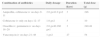
Details of antimicrobial treatment.
| Combination of antibiotics | Daily dosage | Duration (days) | Total dose (g) |
|---|---|---|---|
| Ampicillin, ceftriaxone iv on days 9-11 | 2.0 g×41.0 g×2 | 3 | 246 |
| Ceftriaxone iv only on days 12–17 | 1.0 g×2 | 5 | 10 |
| Oxacillin iv; gentamicin iv on days 18–20 | 2.0 g×40.160 g×1 | 3 | 240.48 |
| Vancomycin iv on days 21–48 | 1 g×2 | 27 | 54 |
iv: intravenous.
S. aureus is the causative agent in the majority of IE cases in IDUs.8 It is the most common pathogen in isolated TV endocarditis, accounting for 50%–60% of cases among IDUs. However, in our case, S. aureus affected not only the TV; vegetations also appeared on the aortic valve (AoV). Less common infectious agents include methicillin-resistant S. aureus (MRSA), Streptococcus bovis and other streptococci species, and fungi.7
Because material is injected intravenously, the first valves to screen particulate matter would be on the right side of the heart. In the present case the patient was addicted to heroin for 10 years. The question arises: why did the patient only develop infective endocarditis to after so many years? Some authors argue that since cocaine has a shorter half-life than heroin it requires more frequent dosing, increasing the bacterial load and incidence of endocarditis.9 According to the literature, if infective endocarditis affects the TV, the prognosis is much more favorable than when it damages the MV or AoV. In the present case the TV was the most seriously damaged valve, but vegetations 6–7 mm in length were also observed on the noncoronary and right coronary leaflets of the AoV, leading to complications involving both sides of the heart. A study by Chambers et al. comparing the clinical presentation of IE among intravenous drug addicts and nonaddicts showed that intravenous drug addicts were younger and had less underlying diseases than nonaddicts.10 In the case presented the patient survived, but according to data in the literature, no other intravenous drug user with so many complications and such a severe condition has survived. There are few clinical studies that analyze the influence of HIV infection in IDUs with IE. Two studies showed that the risk of IE for those infected with HIV is increasing. It is associated with decreased CD4+ cell counts, and among HIV-infected patients with CD4+ cell counts of less than 200 cells/μl or with AIDS criteria, the mortality rate is much higher.6,8 In the present case, despite the patient's diagnosis of HIV infection seven years previously, there was no significant immunosuppression (CD4+ count 651 cells/μl).
Diagnosis of IE is based on the Duke criteria, which are divided into major and minor criteria. A clinical diagnosis of IE requires two major, one major and three minor, or five minor criteria.9 In the case presented, vegetations and regurgitation were detected on transthoracic echocardiography and blood cultures were positive for a typical organism, S. aureus (two major criteria), and the patient was an intravenous drug user (one minor criterion). Due to the presence of two major Duke criteria, a clinical diagnosis was made of infective endocarditis. The key echocardiographic finding in right-sided endocarditis is that of vegetation(s) in association with the TV or rarely the pulmonary valve. Tricuspid vegetations are often large, in excess of 2 cm.11 In the case presented, transesophageal echocardiography was not performed, since transthoracic echocardiography showed vegetations on the TV and AoV, the largest being 29 mm. Diagnosis of IE takes into consideration such indicators as increased ESR and C-reactive protein, splenic enlargement and microscopic hematuria. In our case, CRP of 38 mg/l, ESR of 45 mm/h and hematuria (800/ml) were detected.
Most patients have a benign presentation and the main concern for treatment will be the choice of appropriate antibiotics. This essentially depends on the likely microorganisms, the valves involved and the types of injected drugs the patient has used. In our case only treatment with vancomycin was effective, after which the patient's condition began to improve rapidly.
ConclusionsA severe and exceptional clinical case was presented. The patient was an intravenous drug user, had IE, was HIV-infected, and had damaged TV and AoV. Heart, respiratory and renal failure were rapidly progressing. Despite additional complications, the patient's condition improved significantly after administration of vancomycin, with a significant recovery of EF (from 10% to 50%). Although the patient was given ceftriaxone, oxacillin and gentamicin, only vancomycin produced a good result. It is important to note that even in a patient with a such severe condition, immunosuppression was not detected (CD4+ count 651 cells/μl). The patient was relatively young, and was therefore more likely to survive.
Ethical disclosuresProtection of human and animal subjectsThe authors declare that no experiments were performed on humans or animals for this study.
Confidentiality of dataThe authors declare that they have followed the protocols of their work center on the publication of patient data.
Right to privacy and informed consentThe authors have obtained the written informed consent of the patients or subjects mentioned in the article. The corresponding author is in possession of this document.
Conflict of interestThe authors have no conflicts of interest to declare.
