Informação da revista
Vol. 39. Núm. 9.
Páginas 547-549 (Setembro 2020)
Partilhar
Partilhar
Baixar PDF
Mais opções do artigo
Vol. 39. Núm. 9.
Páginas 547-549 (Setembro 2020)
Image in Cardiology
Open Access
Iatrogenic dissection of the ascending aorta after transcatheter aortic valve implantation treated conservatively
Dissecção da aorta ascendente iatrogénica após implantação de TAVI tratada de forma conservadora
Visitas
3149
Pedro G. Magalhãesa,b,c,
Autor para correspondência
pedrogouveiamagalhaes@gmail.com

Corresponding author.
, Pieter R. Stellab, Michiel Voskuilb, Adriaan O. Kraaijeveldb
a Cardiology Department, Centro Hospitalar de Trás-os-Montes e Alto Douro, Hospital of Vila Real, Vila Real, Portugal
b Interventional Cardiology Department, Division Heart & Lungs, University Medical Center Utrecht (UMCU), Utrecht, Netherlands
c Sociedade Portuguesa de Cardiologia fellow for interventional cardiology fellowship abroad
Este item recebeu

Under a Creative Commons license
Informação do artigo
Texto Completo
Baixar PDF
Estatísticas
Figuras (4)
Mostrar maisMostrar menos
Texto Completo

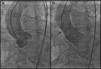
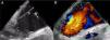
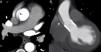
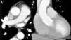
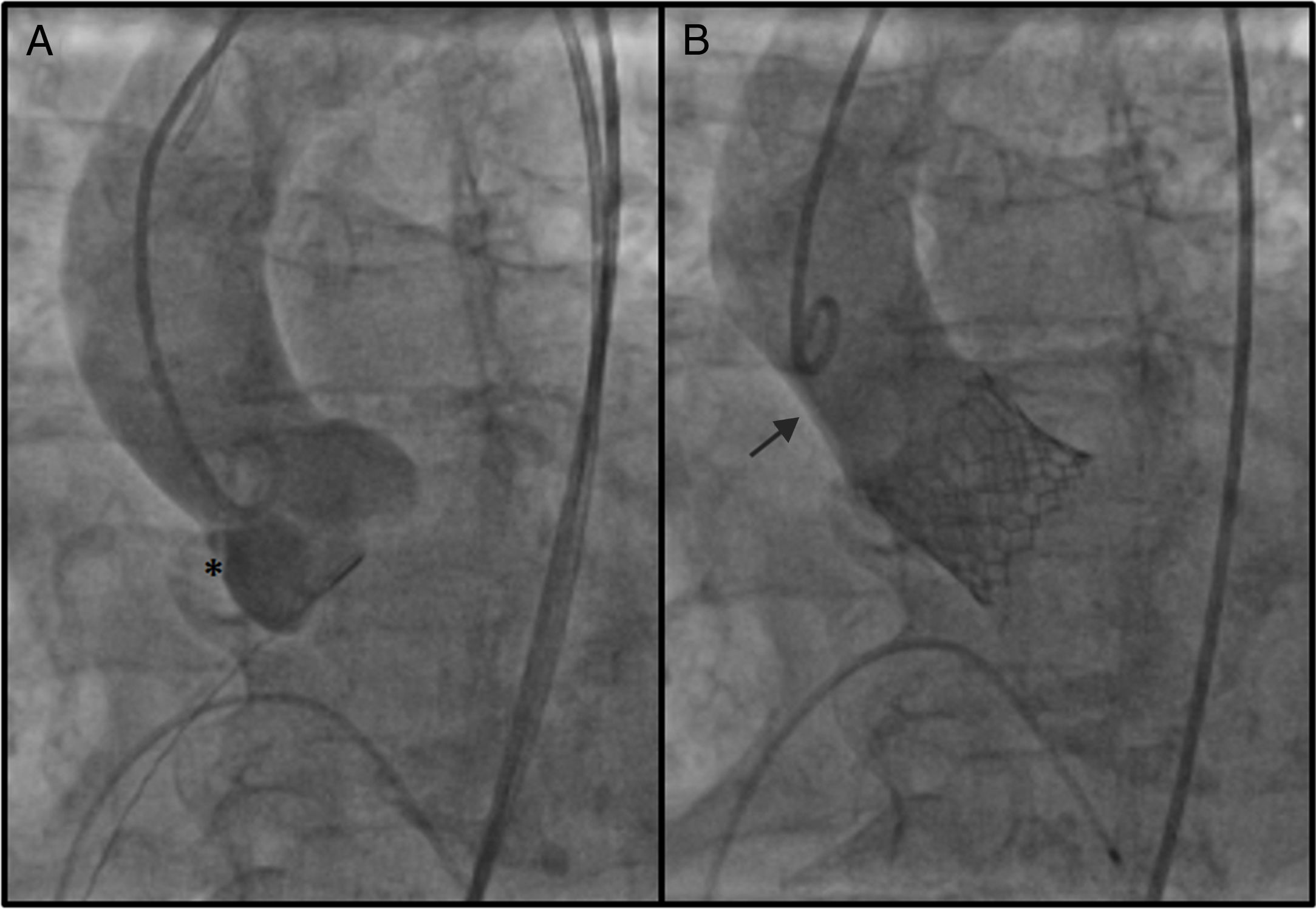
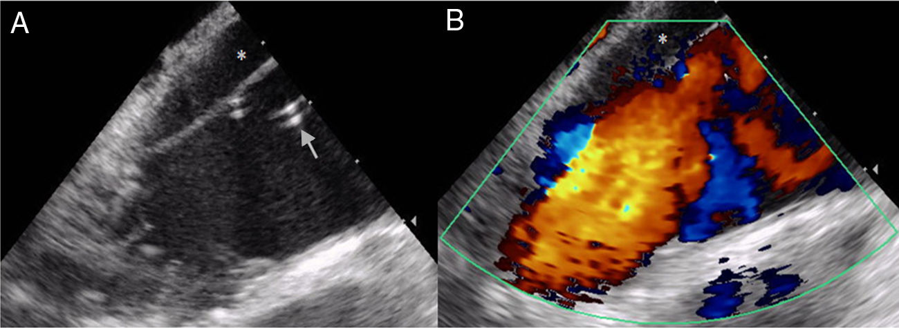
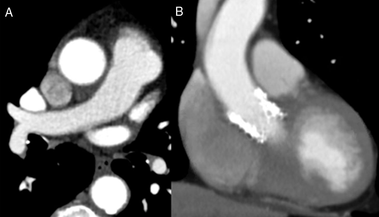
A 90-year-old female with severe symptomatic aortic stenosis was admitted for transcatheter aortic valve implantation (TAVI). A 26-mm Edwards SAPIEN valve was implanted through a transfemoral approach. After the final aortic angiogram a subtraction image was noted from the aortic root just above the right coronary sinus (RCS) to the mid ascending aorta (Figure 1). The intracardiac echocardiogram suggested an aortic dissection, showing an enlarged ascending aorta with an intimal flap without clear flow in the false lumen (Figure 2). The hypothesis of dissection caused by a small tear at the level of the RCS was confirmed by computed tomography (CT) (Figure 3). After heart team discussion it was decided to treat the complication conservatively, since the patient had no symptoms, and she was started on labetalol. A transthoracic echocardiogram was performed that showed good valvular performance without paravalvular leakage. The patient was discharged home after two days. Three months later her exercise capacity had improved and the control CT showed resolution of the dissection (Figure 4).

Figure 1.

Aortography images: (A) after balloon valvuloplasty: * marks the probable starting point of the dissection; (B) after valve placement: subtraction image from the aortic root above the right coronary sinus to the mid ascending aorta.

(0,18MB).
Figure 2.

Intracardiac echocardiography images: (A) intimal flap dividing the aorta into a true lumen, in which the tip of the pigtail catheter can be seen (arrow), and a false lumen (*); (B) color Doppler with virtually no systolic antegrade flow in the false lumen.

(0,09MB).
Figure 3.

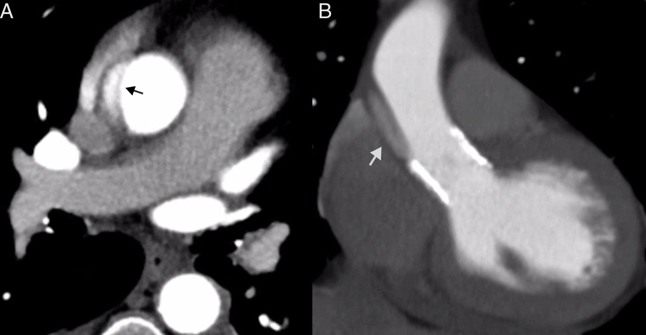
Contrast thoracic computed tomography images: (A) aortic dissection with the intimal tear dividing the aorta into a true lumen, fully contrasted, and a false lumen, only slightly contrasted (arrow); (B) coronal plane reconstruction similar to the aortography view showing the slightly contrasted false lumen (arrow), corresponding to the subtraction image on aortography (Figure 1B).

(0,12MB).
Figure 4.

Control contrast thoracic computed tomography images: complete resolution of the dissection in both transversal (A) and coronal (B) planes.

(0,05MB).

We believe this case highlights the importance of proper assessment of angiographic images that can prompt a search for aortic dissection using other diagnostic means.

Iatrogenic acute aortic dissection complicates 0-1.9% of TAVI cases. The best treatment remains controversial, as mortality in patients treated either with surgery (57-80%) or conservatively (58% at 30 days) is very high. Particularly in very elderly patients, a watchful waiting strategy may be reasonable, as in the case presented here.

Conflicts of interest

The authors have no conflicts of interest to declare.

Idiomas
Revista Portuguesa de Cardiologia
Opções de artigo
Ferramentas
en pt

Are you a health professional able to prescribe or dispense drugs?

Você é um profissional de saúde habilitado a prescrever ou dispensar medicamentos

Ao assinalar que é «Profissional de Saúde», declara conhecer e aceitar que a responsável pelo tratamento dos dados pessoais dos utilizadores da página de internet da Revista Portuguesa de Cardiologia (RPC), é esta entidade, com sede no Campo Grande, n.º 28, 13.º, 1700-093 Lisboa, com os telefones 217 970 685 e 217 817 630, fax 217 931 095 e com o endereço de correio eletrónico revista@spc.pt. Declaro para todos os fins, que assumo inteira responsabilidade pela veracidade e exatidão da afirmação aqui fornecida.