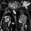
A 64-year-old woman was referred for evaluation of constitutional symptoms, dyspnea and right heart failure. There was a high level of inflammatory markers in the blood test, so blood culture and image tests were performed to rule out endocarditis. Echocardiography did not show valve disease and revealed normal systolic function and a mass at the auriculoventricular sulcus (Figure 1A). Thoracic-abdominal computed tomography ruled out inflammatory and neoplastic diseases, showing the heart mass, perirenal and retroperitoneal fibrosis spreading to pararenal and paraaortic space (Figure 1B) and areas of bone sclerosis in pelvis and vertebrae. Magnetic resonance imaging was performed afterwards. Cardiac cine-balanced fast field echo shows the cardiac mass as an extensive hypointense tissue surrounding the right atrium (Figure 1C) with intense enhancement after intravenous administration of contrast at the inversion-recovery sequences (Figure 1D and E). Intense enhancement was also seen in all aortic wall, supra-aortic trunks, visceral and pulmonary arteries suggesting extensive vascular adventitial fibrosis (Figure 1E). Finally, a retroperitoneal biopsy showed chronic IgG4-lymphoplasmacytic inflammation with a xanthogranulomatous component. In the molecular test, a specific mutation in the BRAF gene was found pointing to Erdheim-Chester disease as the diagnosis.
A: Ecocardiography showing mass at the auriculoventricular sulcus; B: Perirenal and retroperitoneal fibrosis seen in computed tomography; C: Magnetic resonance imaging (MRI) showing cardiac mass and hypointense tissue surrounding right atrium; D and E: MRI, inversion-recovery sequences showing intense enhancement in heart mass and aortic wall.
Erdheim-Chester disease is a rare non-Langerhans histiocytic multisystem disorder (fewer than 1000 cases in the literature) that principally affects long bones (95%), the retroperitoneum (59%) and the cardiovascular system (57%), which can include pseudotumoral cardiac infiltration, valve abnormalities, rhythm defects and periaortic fibrosis. Historically, the prognosis has been poor but new treatments targeted at specific mutations (BRAF inhibitors) may improve survival.
Conflicts of interestThe authors have no conflicts of interest to declare.


