
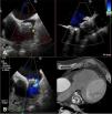

We present the case of an 85-year-old male patient with a history of digestive hemorrhage with hypovolemic shock and hemorrhagic stroke with ad integrum restitution. He had permanent atrial fibrillation (CHA2DS1-VASc: 6 and HAS-BLED: 5) and was receiving indefinite oral anticoagulation with Apixaban®. After the last episode of bleeding, the patient was referred for left atrial appendage closure and underwent a successful 24 mm Watchman device implantation (Boston Scientific, MN, USA), with the presence of a small leak <3 mm that was managed conservatively. Two years after the implant, he suffered a new episode of ischemic stroke in the frontal lobe. Transesophageal echocardiography (TEE) revealed the presence of two significant leaks >5 mm, one on the posterosuperior side and the other on the anteroinferior side (Figure 1, Video 1-2). Due to high hemorrhagic risk, the patient underwent percutaneous closure of the leaks.
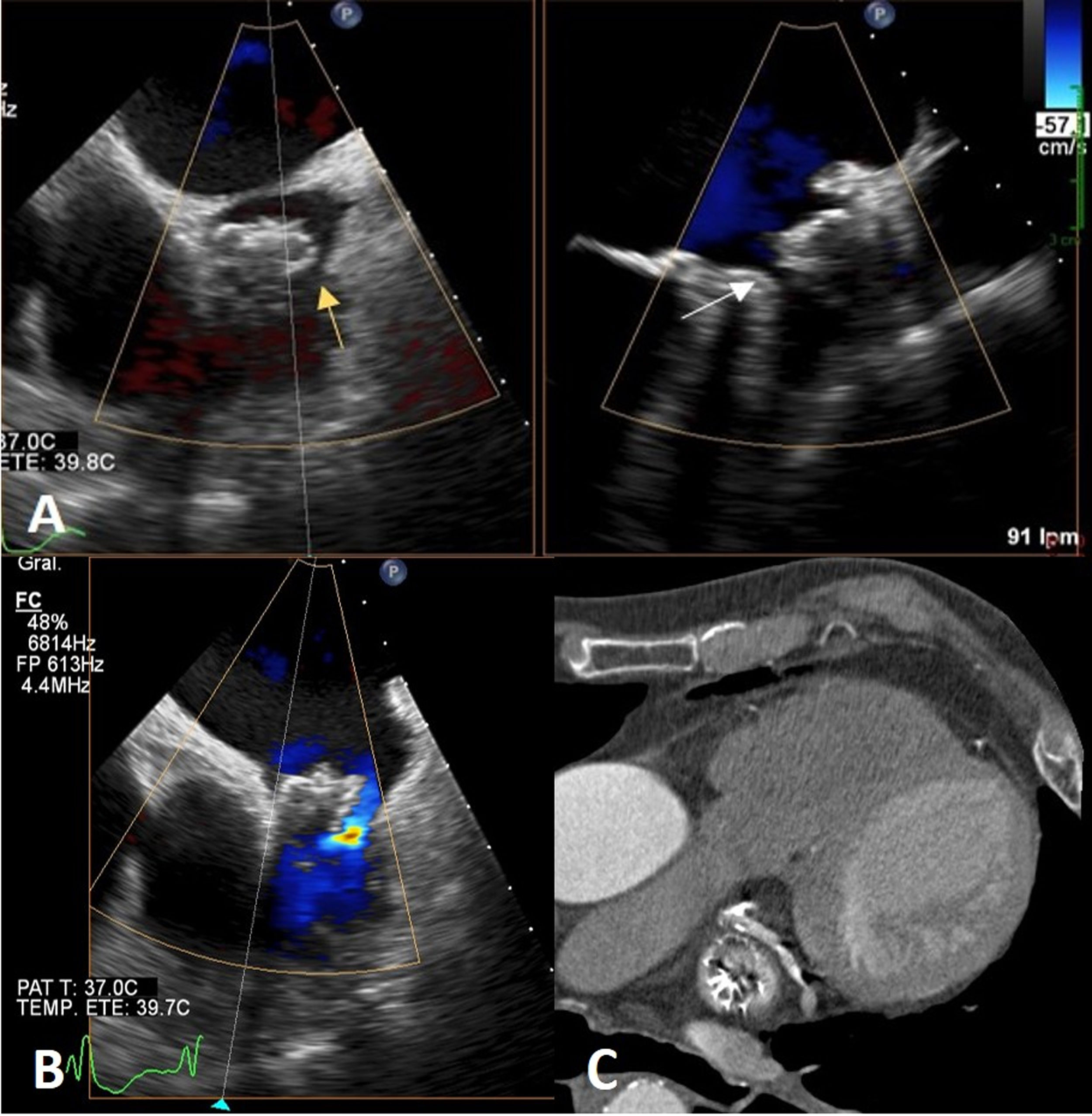
A: Transesophageal echocardiography (TEE) image showing anteroinferior leak (yellow arrow) and posterosuperior leak (white arrow). B: Transesophageal echocardiography with TEE image showing color flow Doppler through the posterosuperior leak. C: Computed tomography image showing leaks on both sides of the device.
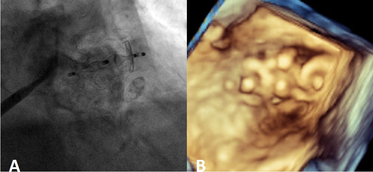
Via right femoral venous access and guided by TEE, transseptal puncture and introduction of a deflectable sheath Agilis 8.5 F Medium Curl (Abbott, IL, USA) in the left atrium were performed. A multipurpose guiding catheter 5F and straight hydrophilic guide (Terumo Europe, Leuven, BE) were introduced through it. Both defects were crossed sequentially and two 12 mm Amplatzer Vascular Plug II (Abbott, IL, USA) devices were released (Figures 2 and 3A and Video 3-6).

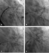
A: Fluoroscopy imaging showing how the guide catheter crossed the upper leak. B: Angiographic image showing the deployment of the first Amplatzer Vascular Plug II into the upper leak. C: Angiographic image showing how the guide catheter crossed the lower leak. D: Fluoroscopy image showing the release of the second Amplatzer Vascular Plug II into the lower leak.
The procedure was performed without complications, and the patient was discharged the following day with aspirin and clopidogrel. The four-week follow-up TEE showed the devices had been implanted appropriately implanted (Figure 3B).
Conflicts of interestThe authors have no conflicts of interest to declare.
