


Cardiac rehabilitation (CR) has been demonstrated to improve exercise capacity in acute coronary syndrome (ACS), but not all patients derive the same benefit. Careful patient selection is crucial to maximize resources.
ObjectiveTo identify in a heterogeneous ACS population which patients would benefit the most with CR, in terms of functional capacity (FC), by using cardiopulmonary exercise testing (CPET).
MethodsA retrospective analysis of consecutive ACS patients who underwent CR and CPET was undertaken. CPET was performed at baseline and after 36 sessions of exercise. Peak oxygen uptake (pVO2), percentage of predicted pVO2, minute ventilation/CO2 production (VE/VCO2) slope, VE/VCO2 slope/pVO2 and peak circulatory power (PCP) (pVO2 times peak systolic blood pressure) were assessed in two moments. The differences in pVO2 (ΔpVO2), %pVO2, PCP and exercise test duration were calculated. Patients were classified according to baseline pVO2 (group 1, <20 ml/kg/min vs. group 2, ≥20 ml/kg/min) and left ventricular ejection fraction (group A, <50% vs. group B, ≥50%).
ResultsWe analyzed 129 patients, 86% male, mean age 56.3±9.8 years. Both group 1 (n=31) and group 2 (n=98) showed significant improvement in FC after CR, with a more significant increase in pVO2, in group 1 (ΔpVO2 4.4±7.3 vs. 1.6±5.4; p=0.018). Significant improvement was observed in CPET parameters in group A (n=34) and group B (n=95), particularly in pVO2 and test duration.
ConclusionPatients with lower baseline pVO2 (<20 ml/kg/min) presented more significant improvement in FC after CR. CPET which is not routinely used in assessement before CR in context of ACS, could be a valuable tool to identify patients who will benefit the most.
A reabilitação cardíaca (RC) melhora a capacidade funcional (CF) de exercício na síndrome coronária aguda (SCA), contudo nem todos os doentes apresentam benefício idêntico. A criteriosa seleção de doentes é crucial para a rentabilização de recursos.
ObjetivoIdentificar na população heterogénea de SCA quais os doentes que mais poderiam beneficiar em termos de capacidade funcional (CF) após programa de RC, através de prova de esforço cardiorrespiratória (PECR).
MétodosRealizámos uma análise retrospetiva de doentes consecutivos com SCA que foram submetidos a RC e PECR. A PECR foi realizada previamente e após 36 sessões de treino de exercício. Parâmetros avaliados: consumo de oxigénio no pico (pVO2), percentagem do pVO2 previsto (%pVO2), declive ventilação minuto/CO2 produzido (VE/VCO2), declive VE/VCO2/pVO2, potência circulatória no pico (PCP) (pVO2 x pressão arterial sistólica no pico). Foram calculadas as variações de pVO2 (ΔpVO2). Os doentes foram analisados de acordo com o pVO2 basal (G1<20 versus G2≥20 ml/kg/min) e fração de ejeção do ventrículo esquerdo (FEVE) (GA<50% versus GB≥50%).
ResultadosAnalisámos 129 doentes, 86% homens, idade média 56,3±9,8 anos. Ambos, G1 (n=31) e G2 (n=98) apresentaram melhoria significativa na CF após RC, com maior aumento significativo de pVO2 no G1 (ΔpVO2 4,4±7,3 versus 1,6±5,4; p=0,018). Verificou-se melhoria dos parâmetros da PECR em GA (n=34) e GB (n=95), particularmente no pVO2 e duração da PECR.
ConclusãoDoentes com menores pVO2 (<20 ml/kg/min) apresentaram, após programa de RC, melhoria mais significativa da CF. A PECR, que não é rotina na avaliação de SCA pré RC, poderá constituir um instrumento valioso na identificação dos doentes que mais beneficiam de RC.
percentage of predicted pVO2
acute coronary syndrome
cardiopulmonary exercise testing
cardiac rehabilitation
exercise training
left ventricular
left ventricular ejection fraction
peak circulatory power
peak oxygen uptake
minute ventilation/CO2 production
Cardiac rehabilitation (CR) has long been considered a cornerstone in secondary prevention after acute coronary syndrome (ACS). It is a coordinated multidisciplinary intervention that aims to improve the cardiac patient's physical, psychological and social functioning. CR programs include exercise training and strategies to reduce modifiable cardiovascular risk factors such as hypertension, dyslipidemia, diabetes and smoking, and to improve adherence to pharmacologic and non-pharmacologic therapy.1,2
CR has been shown to reduce reinfarction, cardiovascular and non-cardiovascular readmission and death3,4,2,5 and to improve health-related quality of life, and to be cost-effective.1,6
Exercise training programs increase patients’ cardiorespiratory fitness, reducing symptoms and improving physiologic responses to physical effort.7 As a consequence, heart rate and blood pressure decrease during physical activity, reducing myocardial oxygen demand.1
The positive effects of CR in cardiovascular disease include stabilization of the progression of atherosclerosis and improvement in endothelial function, resulting in increased coronary compliance, elasticity and flow.1,8–11
Exercise testing is essential before CR to assess exercise capacity, to prescribe exercise training programs, to stratify cardiovascular risk and to estimate prognosis. After CR, it provides an objective measure of the program's effect on functional capacity.
Achievement of higher functional capacity is clinically significant because it not only impacts on health-related quality of life but it also is prognostically valuable, strongly predicting the risk of death. Assessing fitness at baseline and after CR is important since an improvement in functional capacity lowers the risk of death.12
Cardiopulmonary exercise testing (CPET) is the best tool for this purpose, providing a detailed assessment of functional capacity through the analysis of respiratory gas exchange during exercise. Among important CPET parameters is peak oxygen uptake (pVO2), which is an accurate measure of exercise capacity and an important predictor of outcome in patients with coronary artery disease (CAD).13–15 Peak VO2 reflects tissue oxygen uptake in exercise, and depends on cardiac output, arterial oxygen content and oxygen extraction ability of muscle tissue. A supervised exercise training program can increase pVO2 by 11-36%, with the greatest improvement in the most deconditioned individuals.3 Peak VO2 varies according to age and gender, so it is also adjusted in relation to the predicted value for age and gender (%pVO2).15
Another useful parameter, considered by some authors to be a better prognostic parameter than pVO2, is the slope of the ventilation/CO2 ratio (VE/VCO2 slope), which expresses ventilatory efficiency and has been shown to be an independent predictor of outcome.14,16,17 The ratio (VE/VCO2 slope)/pVO2 integrates the two above parameters and also has prognostic value.18
Another form of assessment is calculation of peak circulatory power (PCP), defined as pVO2 times systolic pressure at peak exercise.
Despite its advantages, CPET is not routinely used after ACS.
The aim of the present study was, using CPET parameters, to identify which patients would benefit most in terms of functional capacity after a CR program. Patients were divided according to pVO2 and left ventricular ejection fraction (LVEF) at baseline, and the benefit of CR was evaluated by comparing CPET performance before and after the program.
MethodsStudy designThis was a retrospective observational study of consecutive patients who suffered an ACS and participated in CR after hospital discharge, in whom CPET was performed before and after the program. All patients admitted with ACS were invited to take part in a CR program.
The data were collected between January 2004 and December 2013, in a single center.
The study population was divided into two groups according to baseline functional capacity based on the Weber-Janicki classification19: group 1 (G1), pVO2 <20 ml/kg/min; and group 2 (G2), pVO2 ≥20 ml/kg/min. A second analysis was performed, dividing patients according to left ventricular (LV) systolic function before CR, using the cut-point of 50% LVEF: group A (GA), LVEF <50%; and group B (GB), LVEF ≥50%.
Cardiac rehabilitationEarly ambulatory phase (phase 2) of CR program was performed by a multidisciplinary team, led by a cardiologist.
Before starting the program, patients were stratified according to CAD risk, exercise capacity and safety concerns. All underwent clinical and echocardiographic assessment and exercise testing.
The exercise program was prescribed on the basis of CPET results and was individualized for each patient based on pVO2, in accordance with current guidelines.20
Continuous moderate-intensity exercise training was performed in 36 twice-weekly sessions of 60 min each. Each session included warm-up (10 min), aerobic activity (30 min), muscle-strengthening activity and flexibility exercises (10 min) and cool-down (10 min).
The program was begun at lower intensity (65-70% of maximum training heart rate), and was progressively elevated to a target of 85-95% of maximum training heart rate, without reaching the anaerobic threshold.
Besides the exercise training component, the phase 2 CR program also included:
- –
identification and control of cardiovascular risk factors, including diabetes, hypertension, dyslipidemia, obesity, smoking, and sedentary lifestyle;
- –
lifestyle modification with individualized dietary counseling by a dietician, psychologic counseling for selected patients, smoking cessation sessions for some patients, social support in selected cases and promotion of adherence to drug therapy;
- –
group educational sessions by the multidisciplinary CR team, with the participation of family members, to explain atherosclerotic disease and its consequences and the importance of risk factors and secondary prevention measures.
Immediately after the end of the program, clinical and echocardiographic assessment and exercise testing were repeated, and patients were scheduled for consultations to quantify the benefits obtained the program. Follow-up by telephone was performed to check adherence to healthy behaviors, particularly exercise and medication, control of cardiovascular risk factors and the occurrence of cardiac events.
Cardiopulmonary exercise testingAll patients underwent CPET at baseline and after 36 exercise training sessions.
Maximal symptom-limited treadmill CPET was performed using the modified Bruce protocol. CPET was monitored with continuous 12-lead electrocardiogram, blood pressure cuff, saturation probe and face mask to measure respiratory gases. Blood pressure was measured at rest, at each stage, at peak exercise and at the first, third and sixth minute of the recovery phase. Respiratory gases were analyzed using an Innocor® gas analyzer and VO2, CO2 production (VCO2) and ventilation (VE) were measured on a breath-by-breath basis.
Patients were encouraged to continue exercising until the VCO2/VO2 ratio was ≥1.15.
Peak VO2 adjusted for body mass, or for fat-free mass in obese patients (body mass index >30 kg/m2), was analyzed as %pVO2 according to age and gender. The VE/VCO2 slope was calculated by automatic linear regression with values obtained during the exercise test. The VE/VCO2 slope/pVO2 ratio was calculated using the obtained VE/VCO2 slope and pVO2. PCP was calculated as pVO2 times peak systolic blood pressure. The differences in pVO2, %pVO2, PCP and exercise test duration before and after CR were also calculated.
EchocardiographyAll patients underwent a comprehensive Doppler echocardiographic study using commercially available ultrasound systems (Vivid 9®, General Electric) before and after the CR program.
Echocardiographic parameters of LV systolic and diastolic function were assessed, as well as right ventricular function parameters, pulmonary artery systolic pressure and the presence of cardiac valve disease. LV end-diastolic and end-systolic volume and LVEF were determined by Simpson's biplane method.
Statistical analysisThe statistical analysis was performed using SPSS Statistics (version 22; IBM SPSS, Chicago, IL). Continuous variables were expressed as mean ± standard deviation. CPET parameters at baseline and after CR were compared using the two-tailed Student's t test or the non-parametric Wilcoxon test, as appropriate. Characteristics of the study groups were compared using the Student's t test or the Wilcoxon-Mann-Whitney test for continuous variables and Pearson's chi-square or Fisher's exact test for categorical variables.
ResultsDuring the study period 4724 patients were admitted with ACS diagnosis. Of these, around 50% were transferred from non-tertiary centers without a catheterization laboratory and were followed in those hospitals after discharge. Of the 2363 (50%) followed in our center, only 12% agreed to be included in the hospital's CR program and completed the program.
The study included the 129 patients who underwent CPET before and after CR, 111 (86.0%) male, mean age 56.3±9.8 years, 88.1% of whom had suffered STEMI. The clinical characteristics of the overall population are presented in Table 1.
Clinical characteristics of the overall population and comparison between study groups.
| Overall (n=129) | G1 (n=31) | G2 (n=98) | p | GA (n=34) | GB (n=95) | p | |
|---|---|---|---|---|---|---|---|
| Age (years) | 56.3±9.8 | 61.3±7.8 | 54.8±9.9 | 0.001 | 57.3±10.5 | 56.0±9.6 | 0.497 |
| Male | 86.00% | 74.20% | 89.80% | 0.039 | 94.10% | 83.20% | 0.153 |
| Hypertension | 52.00% | 53.30% | 51.50% | 0.864 | 52.90% | 51.60% | 0.894 |
| Diabetes | 14.20% | 25.80% | 10.40% | 0.042 | 29.40% | 8.60% | 0.007 |
| Dyslipidemia | 59.10% | 80.00% | 52.60% | 0.008 | 55.90% | 60.20% | 0.66 |
| Smoking | 46.10% | 48.40% | 45.40% | 0.769 | 35.30% | 50.00% | 0.14 |
| BMI | 26.8±5.7 | 26.5±6.5 | 26.9±5.5 | 0.837 | 25.9±79 | 27.2±4.8 | 0.898 |
| Family history of CAD | 14.80% | 9.70% | 16.50% | 0.562 | 11.80% | 16.00% | 0.556 |
| STEMI | 88.10% | 90.00% | 87.50% | 0.864 | 88.20% | 88.00% | 0.497 |
| No. of vessels with CAD | 1.5±0.7 | 1.6±0.8 | 1.5±0.6 | 0.626 | 1.6±0.7 | 1.5±0.6 | 0.578 |
| Untreated CAD | 26.40% | 26.70% | 26.30% | 0.97 | 26.50% | 26.40% | 0.991 |
| LVEF (%) | 54.1±9.9 | 53.4±11.9 | 54.3±9.2 | 0.662 | 41.9±7.3 | 58.5±6.4 | <0.001 |
| Sinus rhythm | 90.60% | 93.50% | 89.70% | 0.729 | 85.30% | 92.60% | 0.3 |
BMI: body mass index; CAD: coronary artery disease; G1: group 1 (pVO2 <20 ml/kg/min); G2: group 2 (pVO2 ≥20 ml/kg/min); GA: group A (LVEF <50%); GB: group B (LVEF ≥50%); LVEF: left ventricular ejection fraction; STEMI: ST-segment myocardial infarction.
Compared to G1, G2 patients (with lower baseline functional capacity) were older, included fewer males and had a higher prevalence of diabetes and dyslipidemia (Table 1).
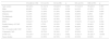
CR improved functional capacity in both groups. In G1, pVO2 increased from 17.7±1.8 to 22.1±7.2 ml/kg/min (p<0.001). PCP and exercise test duration also increased significantly (p<0.001 for both). VE/VCO2 slope/pVO2 decreased from 1.7±0.4 to 1.4±0.6 (p<0.001). There was no significant change in VE/VCO2 slope in this group (Table 2).
Comparison of cardiopulmonary exercise testing parameters before and after cardiac rehabilitation in the study groups.
| CPET parameters | G1 (n=31) | G2 (n=98) | GA (n=34) | GB (n=95) | ||||||||
|---|---|---|---|---|---|---|---|---|---|---|---|---|
| Before CR | After CR | p | Before CR | After CR | p | Before CR | After CR | p | Before CR | After CR | p | |
| Peak VO2 (ml/kg/min) | 17.7±1.8 | 22.1±7.2 | <0.001 | 28.1±5.6 | 29.7±6.2 | <0.001 | 24.7±6.1 | 27.7±6.9 | 0.002 | 26.0±6.9 | 28.0±7.3 | 0.001 |
| % predicted peak VO2 | 68.6±12.3 | 86.5±33.0 | <0.001 | 96.3±21.7 | 100.3±21.8 | 0.001 | 84.9±19.4 | 95.4±24.0 | 0.001 | 91.4±24.1 | 97.6±26.1 | 0.003 |
| VE/VCO2 slope | 29.2±6.1 | 28.6±5.8 | 0.213 | 25.7±5.5 | 24.9±4.6 | 0.031 | 25.9±5.4 | 25.5±4.9 | 0.241 | 26.8±5.9 | 26.0±5.2 | 0.028 |
| PCP (mmHg.ml/kg/min) | 2863.7±581.6 | 4866.4±1213.0 | <0.001 | 4866.4±1213.0 | 5104.9±1196.3 | <0.001 | 4206.7±1372.4 | 4583.8±1417.4 | 0.034 | 4449.0±1398.0 | 4848.3±1367.2 | <0.001 |
| (VE/VCO2 slope)/pVO2 | 1.7±0.4 | 1.4±0.6 | <0.001 | 1.0±0.3 | 0.9±0.3 | 0.031 | 1.1±0.5 | 1.0±0.6 | 0.003 | 1.1±0.4 | 1.0±0.4 | <0.001 |
| Duration (min) | 10.8±2.6 | 13.2±2.4 | <0.001 | 14.2±2.8 | 15.9±2.5 | <0.001 | 13.4±2.6 | 15.0±3.3 | <0.001 | 13.4±3.3 | 15.4±2.5 | <0.001 |
CPET: cardiopulmonary exercise testing; G1: group 1 (pVO2 <20 ml/kg/min); G2: group 2 (pVO2 ≥20 ml/kg/min); GA: group A (LVEF <50%); GB: group B (LVEF ≥50%); PCP: peak circulatory power.
In G2 all CPET parameters improved after CR, pVO2 rising from 28.1±5.6 to 29.7±6.2 ml/kg/min (p<0.001) and %pVO2 from 96.3% to 100.3% (p=0.001). PCP and exercise test duration also increased significantly. VE/VCO2 slope and VE/VCO2 slope/pVO2 decreased significantly (p=0.032 and p<0.001, respectively) (Table 2).
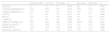
As expected, CPET parameters were substantially different between groups before and after CR, with a better performance in G2. However, when range of improvement is compared, G1 achieved a greater benefit after CR. The improvement in pVO2 was greater in G1, in absolute value (ΔpVO2 4.4±7.3 vs. 1.6±5.4; p=0.018) and in percentage of predicted value (Δ%pVO2 17.9±35.4% vs. 4.0±19.9%; p=0.009). In G1 improvement in pVO2 was more than 10% in 71% of patients, contrasting with only 40.8% in G2. The increase in PCP was also significantly greater in G1 (883.3±1217.3 vs. 238.5±1250.5 mmHg.ml/kg/min; p=0.015). Exercise test duration increased more in G1, although not significantly (Table 3).
Comparison between cardiopulmonary exercise testing parameters before and after cardiac rehabilitation in groups 1 and 2.
| CPET parameters | G1 (n=31) | G2 (n=98) | p |
|---|---|---|---|
| Peak VO2 (ml/kg/min) before CR | 17.7±1.8 | 28.1±5.6 | <0.001 |
| Peak VO2 (ml/kg/min) after CR | 22.1±7.2 | 29.7±6.2 | <0.001 |
| % predicted peak VO2 before CR | 68.6±12.3 | 96.3±21.7 | <0.001 |
| % predicted peak VO2 after CR | 86.5±33.0 | 100.3±21.8 | <0.001 |
| VE/VCO2 slope before CR | 29.2±6.1 | 25.7±5.5 | 0.003 |
| VE/VCO2 slope after CR | 28.6±5.8 | 24.9±4.6 | 0.002 |
| PCP (mmHg.ml/kg/min) before CR | 2863.7±581.6 | 4866.4±1213.0 | <0.001 |
| PCP (mmHg.ml/kg/min) after CR | 3746.9±1432.0 | 5104.9±1196.3 | <0.001 |
| (VE/VCO2 slope)/pVO2 before CR | 1.7±0.4 | 1.0±0.3 | <0.001 |
| (VE/VCO2 slope)/pVO2 after CR | 1.4±0.6 | 0.9±0.39 | <0.001 |
| Duration (min) before CR | 10.8±2.6 | 14.2±2.8 | <0.001 |
| Duration (min) after CR | 13.2±2.4 | 15.9±2.5 | <0.001 |
| ΔpVO2 | 4.4±7.3 | 1.6±5.4 | 0.018 |
| Δ%pVO2 | 17.9±35.4 | 4.0±19.9 | 0.009 |
| Increase in pVO2 >10% | 71.0% | 40.8% | 0.003 |
| Δ CPET duration (min) | 2.5±2.8 | 1.7±2.5 | 0.101 |
| Δ PCP (mmHg.ml/kg/min) | 883.3±1217.3 | 238.5±1250.5 | 0.015 |
Δ: difference; CPET: cardiopulmonary exercise test; CR: cardiac rehabilitation; G1: group 1 (pVO2 <20 ml/kg/min); G2: group 2 (pVO2 ≥20 ml/kg/min); PCP: peak circulatory power.
When patients were grouped according to LV systolic function, both groups presented improved functional capacity after exercise training. In GA, pVO2 increased from 24.7±6.1 to 27.7±6.9 ml/kg/min (p=0.002), %pVO2 from 84.9±19.4 to 95.4±24.0% (p=0.001) and exercise test duration from 13.4±2.6 to 15.0±3.3 min (p<0.001) (Table 2). GB patients also achieved a significant improvement, as demonstrated by increased pVO2, %pVO2, PCP and exercise test duration and decreased VE/VCO2 slope and (VE/VCO2 slope)/pVO2 ratio (Table 2).
There were no significant differences in CPET parameters between these two groups before or after CR. GA presented greater increases in pVO2 (3.1±5.6 vs. 2.0±6.1 ml/kg/min; p=0.382), %pVO2 (10.6±19.0 vs. 6.2±26.9%; p=0.291) and exercise test duration (1.6±2.5 vs. 2.0±2.6; p=0.920), although they were not significant.
In patients with initial VE/VCO2 slope >30, a significant improvement in this parameter was achieved after CR (34.4±5.8 vs. 31.1±5.1; p=0.02).
In a follow-up of 5.17±2.04 years, there were no significant differences in death, cardiac events, lifestyle or adherence to therapy between the study groups (Table 4). The lower mortality and ACS recurrence in all groups is noteworthy. Regarding lifestyle, more than 90% of the overall population maintained therapeutic adherence and more than half of patients performed regular physical exercise and kept to a healthy diet.
Comparison of follow-up parameters in the study groups.
| Overall (n=129) | G1 (n=31) | G2 (n=98) | p | GA (n=34) | GB (n=95) | p | |
|---|---|---|---|---|---|---|---|
| Death (%) | 4.7 | 3.2 | 5.1 | 0.555 | 8.0 | 3.8 | 0.329 |
| All-cause hospitalization (%) | 47.9 | 46.7 | 48.3 | 0.876 | 47.8 | 47.9 | 0.994 |
| Cardiac hospitalization (%) | 31.8 | 25.8 | 33.7 | 0.412 | 32.0 | 31.7 | 0.979 |
| ACS (%) | 6.2 | 0.0 | 8.2 | 0.197 | 4.0 | 6.7 | 0.999 |
| PCI (%) | 13.2 | 6.5 | 15.3 | 0.359 | 4.0 | 15.4 | 0.192 |
| CABG (%) | 1.6 | 0.0 | 2.0 | 0.999 | 4.0 | 1.0 | 0.351 |
| Adherence to therapy (%) | 95.6 | 92.9 | 96.5 | 0.595 | 100.0 | 94.6 | 0.581 |
| Regular exercise (%) | 53.5 | 40.9 | 57.8 | 0.170 | 73.3 | 49.3 | 0.090 |
| Healthy diet (%) | 54.4 | 47.6 | 54.1 | 0.608 | 57.1 | 51.5 | 0.699 |
| Risk factor control (%) | 75.0 | 68.0 | 77.2 | 0.354 | 75.0 | 75.0 | 1.000 |
ACS: acute coronary syndrome; CABG: coronary artery bypass grafting; G1: group 1 (pVO2 <20 ml/kg/min); G2: group 2 (pVO2 ≥20 ml/kg/min); GA: group A (LVEF <50%); GB: group B (LVEF ≥50%); PCI: percutaneous coronary intervention.
The main result of our study was that the CPET parameter pVO2 is useful in the selection of ACS patients who will have a better functional response to exercise training.
Our data indicate that the inclusion of ACS patients in a supervised 36-session exercise training program improved functional capacity independently of baseline performance, and that patients with pVO2 <20 ml/kg/min before CR derived greater benefit. The improvement in pVO2 was seen in absolute terms, as well as adjusted to predicted value for age and gender. As demonstrated in previous papers,1,21 our data showed that the most deconditioned individuals had the greatest improvement in functional capacity after the program, with 71% of patients in G1 achieving an improvement of >10% in pVO2 compared to 40.8% in G2.
This greater benefit was also reflected in VE/VCO2 slope/pVO2, PCP and exercise test duration, which have important prognostic implications. Kavanagh et al. demonstrated that the main predictor of outcome in CAD patients is exercise tolerance, as objectively measured by pVO2. A 10% decrease in mortality was observed for each increase of 1 ml/kg/min in pVO2.13
Comparing patients according to baseline LVEF, our research demonstrates that CR is beneficial regardless of LV systolic function. More than half of the patients (55.9%) in GA saw an increase in pVO2 of >10%, compared to 45.3% in GB. This finding, although not statistically significant, suggests that patients with LV dysfunction may derive more benefit from an exercise training program. Previous studies have shown that patients with LV systolic dysfunction benefit more from CR.22
The number of ACS patients undergoing CR was low for various reasons, as analyzed in previous studies, including financial constraints, difficulties in transportation, conflicts with work, low motivation and geographic distance. There are multiple barriers to CR, three of which have been identified as patient-related, physician-related and health system-related factors.23
Cardiac rehabilitation, though widely recommended in ACS guidelines, is poorly implemented in several countries, including Portugal, where previous studies have shown that only 3% of ACS patients discharged from hospital are admitted to a CR program.24–26 Although an increasing number of ACS patients have been included in CR programs in Portugal in recent years, recruitment in the country remains far below the European average of 30%.27
Including more patients in CR programs will require modifications in the health system, including increases in the number of CR centers and improvements in specialized training of health professionals. Failure by cardiologists and surgeons to refer patients for CR is particularly evident in some cases, including the elderly, women, obese patients, smokers and those of low socioeconomic status or suffering from depression.28 Only with a more suitable health structure and better trained professionals will be possible to recruit and motivate more ACS patients for CR.
Although in our center there is presently no limitation on the number of ACS patients who can be referred for CR, it should be borne in mind that many hospitals do not have facilities available to provide CR for all patients. Knowing who will derive the greatest benefit will help in the selection of the best responders to CR.
Study limitationsThis is a single-center retrospective study and thus suffers from inherent limitations, including the small number of patients. The size of our sample was due to the fact that of the patients followed after discharge in our center, only a minority agreed to be included in CR at the hospital and completed the program, for the reasons described.
Furthermore, the present study only included patients who performed CPET before and after CR, and since CPET is not a routine procedure, this excluded patients who underwent standard exercise testing, which does not include gas exchange analysis.
ConclusionIn conclusion, of the heterogeneous group of patients with ACS, those with the worst functional capacity identified by CPET, and those with lower LVEF, obtained the greatest benefit from CR. These individuals should be the main target for referral for comprehensive CR, and CPET may be in fact a valuable tool for their identification.
Ethical disclosuresProtection of human and animal subjectsThe authors declare that no experiments were performed on humans or animals for this study.
Confidentiality of dataThe authors declare that they have followed the protocols of their work center on the publication of patient data.
Right to privacy and informed consentThe authors have obtained the written informed consent of the patients or subjects mentioned in the article. The corresponding author is in possession of this document.
Conflicts of interestThe authors have no conflicts of interest to declare.
