


Cardiac papillary fibroelastomas (CPF) are the second most common primary cardiac tumor with highest prevalence in the sixth to eighth decade of life. They involve predominantly the valves, and 80% arise from the valvular endocardium.
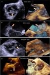
The authors present a transesophageal echocardiography imaging case series including three-dimensional imaging of CPF, confirmed by histopathology, involving the four cardiac valves. White arrow corresponds to cardiac papillary fibroelastomas. Panel A: two dimensional and three dimensional (3D) mid esophageal aortic valve short-axis view showing cardiac papillary fibroelastoma (14x7 mm) at the right coronary aortic cusp in a 73 year-old woman; Panel B: mid-esophageal two chamber view showing a highly mobile and spherical pedunculated tumor (13x13 mm) attached to the posterior mitral leaflet base (white asterisk) in an asymptomatic 60 year-old man; Panel C: Transgastric right ventricle inflow view showing pedunculated and independently mobile tumor mass (13x11 mm) attached to the posterior leaflet of the tricuspid valve (white asterisk) in a 57 year-old woman; Panel D: Mid-esophageal aortic short-axis view showing an unusual and highly mobile tumor mass in pulmonary artery in 42 years old-man, Figure 1.

The panel shows four different patients with cardiac papillary fibroelastomas involving the four cardiac valves. For standardization purposes, the images were obtained during transesophageal echocardiography including three-dimensional imaging which provides complementary anatomical information, identifying the attachment point to the posterior mitral leaflet base and posterior leaflet of the tricuspid valve (white asterisk).
(White arrow corresponds to cardiac papillary fibroelastomas. RA: right atrium; RV: right ventricle; AV: aortic valve; PA: pulmonary artery.)
The images illustrate features that can help establish a differential diagnosis from other intracardiac tumors and masses, such as independent mobility, the presence of papillary fronds similar to a “sea anemone” with a shimmering border, a usual diameter of between 5-40 mm and the presence of a short stalk. As demonstrated in this image case series, the use of three dimensional echocardiography gives additional value, enabling better characterization of CPF anatomy, which is essential in surgical planning.
Conflicts of interestThe authors have no conflicts of interest to declare.
