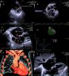
We report the case of a 79-year-old man hospitalized for stroke. The patient had hypertension and dyslipidemia and the ECG revealed previously unknown atrial fibrillation. A transthoracic echocardiogram (TTE) was performed two days after admission to exclude an embolic source. This showed marked dilatation of the right cavities, systolic–diastolic flattening of the interventricular septum (IVS) (Figure 1, panel A) and depressed right ventricular systolic function (S′: 8cm/s; 3D ejection fraction: 18%) (Figure 1, panel D; Video 1). The left ventricle was small and compressed by the bulging IVS; no other wall motion abnormalities were observed. There was a large thrombus 35mm×19mm in size on the right atrium (RA) lateral wall (Figure 1, panel B), and another thrombus 40mm in length through the patent foramen ovale (PFO) (Figure 1, panel C; Video 2).
(A) Transthoracic echocardiography (TTE) image showing marked dilatation of the right chambers with systolic-diastolic flattening of the interventricular septum (IVS); (B) 3D TTE estimation of right ventricularejection fraction; (C) 2D TTE showing a large thrombus onthe right atrium (RA) lateral wall; (D) 2D TTE showing a thrombus through the patentforamen ovale(PFO); (E) Thoracic computed tomography angiographyshowing alarge thrombus onthe RA lateral wall; (F) 2D TTE showing the disappearance of the thrombus through the PFO, after one week of anticoagulation.
The patient underwent thoracic CT angiography that confirmed signs of chronic pulmonary embolism and assessed the larger thrombus described (Figure 1, panel E). Considering the patient's comorbidities, medical treatment was proposed with anticoagulation using heparin. A week later, no thrombus could be observed through the PFO (Figure 1, panel F), although the larger thrombus in the RA was unchanged. The patient was discharged on the 10th day and is recovering uneventfully.
This case highlights the role of TTE in the study of cardioembolic stroke in terms of diagnosis and subsequent treatment strategy. The finding of a large thrombus in the RA and another through the PFO is a rare occurrence; in this case the latter was the cause of both stroke and pulmonary embolism.
Ethical disclosuresProtection of human and animal subjectsThe authors declare that no experiments were performed on humans or animals for this investigation.
Confidentiality of dataThe authors declare that no patient data appear in this article.
Right to privacy and informed consentThe authors declare that no patient data appear in this article.
Conflicts of interestThe authors have no conflicts of interest to declare.


