


A 46-year-old man with no history of cardiovascular disease was admitted with shortness of breath. Clinical examination showed sinus tachycardia (115 bpm) with right bundle branch block and normal blood pressure. Transthoracic echocardiography revealed normal left ventricular size and systolic function, normal right ventricular size and systolic function and normal size of the atria.
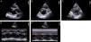
Parasternal long-axis (Figure 1A) and short-axis views (Figure 1B: mitral level, Figure 1C: papillary muscle level) showed a unileaflet mitral valve with an elongated and thickened anterior leaflet, while the posterior leaflet was extremely hypoplastic and almost entirely absent (Video 1). On M-mode imaging of the mitral valve, there was fusion of the anterior mitral E-F and F-A waves, appearing as a single flat E-A wave with loss of the F point (asterisk in Figure 1D) and in contact with the interventricular septum. There were minimal waves (not clearly distinct) of the hypoplastic posterior mitral leaflet and the coaptation zone, depicted by the C point, was posteriorly displaced (Figure 1D). Figure 1E shows the normal M-mode waves of a normal bileaflet mitral valve.
![Transthoracic echocardiography. Two-dimensional parasternal long-axis (A) and short-axis [(B) mitral valve level, (C) papillary muscle level] views; M-mode images at the level of the mitral leaflets of the present unileaflet mitral valve case (D) and of a normal bileaflet mitral valve (E). D point: initial diastolic leaflet opening; E point: maximum leaflet opening during the phase of rapid ventricular filling ending at the F point; A point: maximum leaflet opening during atrial contraction; C point: leaflet coaptation point at the beginning of systole. In the present case of a unileaflet mitral valve (D), there is loss of the F point (asterisk) with a flat E-A wave in constant contact with the anterior interventricular septum, corresponding to the opening of the elongated anterior mitral leaflet throughout diastole, while the coaptation point C is posteriorly displaced.](https://static.elsevier.es/multimedia/08702551/0000004300000012/v1_202412020735/S0870255124001896/v1_202412020735/en/main.assets/gr1.jpeg?xkr=ue/ImdikoIMrsJoerZ+w94UphxYc+GPca8Z7OggvdfJQF4SIqTc4zp8SrbcUWBiK2zRV+4/JKcouWkO4IeVBdVCZ20ei79qgUubvBq8Axg/C8EtmvUPioMBxDcsvj91QqicrBpvptd/54y18nxyNXlQmE4JMSbizD1mUFyENCVqpFhwKdS8EYFbF8QClsyRJVyOu+MmqWJf7SmxaMS4pte7OVumJaNRWxwGixUedbP6EvDrHM3HBvnmiwWYxnSAr1hMocidowWvSbsc3SzvehBcuI13bzu44g76tm8VzY30=)
Transthoracic echocardiography. Two-dimensional parasternal long-axis (A) and short-axis [(B) mitral valve level, (C) papillary muscle level] views; M-mode images at the level of the mitral leaflets of the present unileaflet mitral valve case (D) and of a normal bileaflet mitral valve (E). D point: initial diastolic leaflet opening; E point: maximum leaflet opening during the phase of rapid ventricular filling ending at the F point; A point: maximum leaflet opening during atrial contraction; C point: leaflet coaptation point at the beginning of systole. In the present case of a unileaflet mitral valve (D), there is loss of the F point (asterisk) with a flat E-A wave in constant contact with the anterior interventricular septum, corresponding to the opening of the elongated anterior mitral leaflet throughout diastole, while the coaptation point C is posteriorly displaced.
Doppler imaging revealed mild mitral regurgitation, trivial tricuspid regurgitation and no signs of pulmonary hypertension. The aortic valve was tricuspid and the aortic dimensions were normal. Transesophageal echocardiography confirmed the previous findings (Video 1).
Conflicts of interestThe authors have no conflicts of interest to declare.
