
Bioresorbable vascular scaffolds (BVS) were recently approved for percutaneous coronary intervention in Europe. The aim of this position statement is to review the information and studies on available BVS, to stimulate discussion on their use and to propose guidelines for this treatment option in Portugal.
Methods and ResultsA working group was set up to reach a consensus based on current evidence, discussion of clinical case models and individual experience. The evidence suggests that currently available BVS can produce physiological and clinical improvements in selected patients. There are encouraging data on their durability and long-term safety. Indications were grouped into three categories: (a) consensual and appropriate – young patients, diabetic patients, left anterior descending artery, long lesions and diffuse disease; (b) less consensual but possible – small collateral branches, stabilized acute coronary syndromes; and (c) inappropriate – left main disease, tortuosity, severe calcification.
ConclusionBVS are a viable treatment option based on the encouraging evidence of their applicability and physiological and clinical results. They should be used in appropriate indications and will require technical adaptations. Outcome monitoring and evaluation is essential to avoid inappropriate use. It is recommended that medical societies produce clinical guidelines based on high-quality registries as soon as possible.
Os suportes vasculares restaurativos transitórios (sVRT) foram recentemente aprovados para intervenção coronária percutânea (ICP) na Europa e possuem propriedades muito inovadoras. O objetivo desta declaração de posição é rever criticamente a informação e os estudos com os sVRT disponíveis e contribuir para uma reflexão científica que promova o seu uso racional com orientações estruturadas para a sua aplicação inicial em Portugal.
Métodos e resultadosFoi constituído um grupo de trabalho para alcançar um consenso com base na evidência científica conhecida, na discussão de casos clínicos modelo e na experiência individual. A evidência reunida sugere que os sVRT disponíveis podem produzir uma melhoria fisiológica e clínica em doentes selecionados. Os dados relativos à sua durabilidade e segurança a longo prazo são animadores. As indicações iniciais foram agrupadas em três categorias: a) consensuais e apropriadas – jovens, diabéticos, descendente anterior, lesões longase doença difusa, b) menos consensuais mas possíveis – lesões com pequeno colateral, síndromas coronárias agudas estabilizadas; c) inapropriadas – tronco comum, tortuosidade, calcificação grave.
ConclusãoOs suportes vasculares restaurativos transitórios constituem uma terapêutica válida pela evidência científica encorajadora da sua aplicabilidade, da melhoria fisiológica e clínica. Devemos privilegiar as indicações aconselhadas e adequar as técnicas de angioplastia coronária, bem como monitorizar e avaliar os resultados para evitar uma adoção inapropriada. É recomendável o desenvolvimento expedito de normas de orientação clínica pelas sociedades científicas apoiada em registos de elevada qualidade.
Andreas Gruntzig performed the first coronary balloon angioplasty in 1977,1 and since then there have been continual advances in the percutaneous treatment of coronary artery disease by cardiac catheterization.
A major development occurred in 1986 with the introduction of stents, which reduced the rate of subacute coronary artery occlusion to 1.5%, considerably decreasing the need for emergency coronary artery bypass grafting.2
The next advance was in 2001 with the advent of drug-eluting stents (DES), which, by lessening neointimal hyperplasia, dramatically reduced the restenosis rate seen with bare-metal stents by 39–61%, and hence the need for secondary revascularization.3–5
The introduction of DES sparked a wealth of research on coronary devices that included registries and high-quality randomized trials, contributing to evidence-based medicine in this area. This demonstrated that the increasingly widespread use of DES had limitations, particularly in terms of late thrombosis, which has now been thoroughly studied and controlled.5,6
Despite the good clinical outcomes obtained with DES, these stents have a fixed, rigid metal structure that cannot be removed and that hinders the adaptive biological process of remodeling. Furthermore, the polymers and drugs involved cause local inflammation, which inhibits physiological recovery of the artery and contributes to late thrombosis and neoatherosclerosis.
After a decade of intense pre-clinical research, there was a third revolutionary advance, that of bioresorbable vascular scaffolds (BVS), which are designed to provide temporary radial support to the vessel, to facilitate administration of antiproliferative drugs and to promote recovery of the artery's normal structure and physiological function by gradual removal of the scaffolding through a process of biodegradation.
BVS have several advantages, including physiological recovery of the vessel, reduced stent thrombosis and need for antiplatelet therapy, fewer constraints on future interventions in the vessel and its collaterals, and the possibility of using noninvasive diagnostic exams, particularly computed tomography angiography.7–11 These devices afford all the benefits of a stent, plus the added advantage of being absorbed by the body, ideally after they have fulfilled their function, imposing no constraints on future interventions (Table 1).
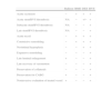
Potential advantages of bioresorbable vascular scaffolds.
| Balloon | BMS | DES | BVS | |
| Acute occlusion | − | + | + | + |
| Acute stent/BVS thrombosis | NA | − | +/− | + |
| Subacute stent/BVS thrombosis | NA | − | − | + |
| Late stent/BVS thrombosis | NA | − | − | + |
| Acute recoil | − | + | + | + |
| Constrictive remodeling | − | + | + | + |
| Neointimal hyperplasia | − | − | + | + |
| Expansive remodeling | − | − | − | + |
| Late luminal enlargement | + | − | − | + |
| Late recovery of vasomotion | − | − | − | + |
| Preservation of collaterals | − | − | − | + |
| Preservation for CABG | + | − | − | + |
| Noninvasive evaluation of treated vessel | + | − | − | + |
BMS: bare-metal stent; CABG: coronary artery bypass grafting; DES: drug-eluting stent; NA: not applicable because of absence of stent; +: prevented or not restricted; −: not prevented or restricted.
BVS systems were recently approved for percutaneous coronary intervention in Europe and possess highly innovative properties. The aim of this position statement is to review the information and studies on available BVS, to stimulate discussion on their use and to propose guidelines for their application by interventional cardiologists in Portugal.
MethodsA working group of experienced interventional cardiologists was set up to evaluate current knowledge and to reach a consensus based on available evidence, discussion of clinical case models and individual experience.
Information and current evidence on bioresorbable vascular scaffolds under developmentBVS have been under development for more than a decade (Table 2).10 The first BVS implanted in humans was the Igaki-Tamai (Igaki Medical Planning Company, Kyoto, Japan) in 2000, with poly-L-lactic acid (PLLA) scaffolds, using a complex thermal delivery technique consisting of balloon inflation with a heated dye at 80°C.12 The first metal BVS used in humans, in 2007, was composed of 93% magnesium (Biotronik, Berlin, Germany),13 while the first coated BVS appeared in 2008 – the everolimus-eluting ABSORB stent (Abbott Vascular, Santa Clara, US) – with a strut thickness of 150 mm.14
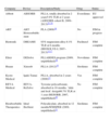
Bioresorbable vascular scaffolds.
| Company | Device | Description/Study | Drug | Status |
| Abbott | ABSORB | PLLA, totally absorbed in 2 years5.9% TLR at 2 years (ABSORB cohort B, 2009–2013)22–24 | Everolimus | EU approved |
| ART | ART Bioresorbable stent | PLA (2009)16 | No | FIM in progress |
| Biotronik | DREAMS | 93% magnesium alloy.9.1% TLR at 6 months (BIOSOLVE-I, 2007–2013)13,17 | Paclitaxel | FIM complete |
| Elixir | DESolve | PLLABDES program (2009, unpublished)18 | Novolimus | FIM complete |
| Huaan | Xinsorb | PLLA (2012)19 | Sirolimus | FIM complete |
| Kyoto Medical | Igaki-Tamai | PLLA, absorbed in 2 years (2000)12 | Yes | FIM complete |
| REVA Medical | REVA-ReZolve | Tyrosine polycarbonate, absorbed in 18 months, ‘slide and lock’ design66.7% TLR at 1 year (RESORB, 2007, unpublished)20 | No | FIM complete |
| Bioabsorbable Therapeutics | Ideal BioStent | Polysalicylate, absorbed in 12 monthsWHISPER (2009, unpublished)21 | Sirolimus | FIM complete |
FIM: first-in-man; PLA: polylactic acid; PLLA: poly-L-lactic acid; TLR: target lesion revascularization.
At present, the ABSORB everolimus-eluting system is the only BVS available for clinical use.
In the ABSORB cohort A trial, with 30 patients, the major event rate at four years was only 3.4%, demonstrating that the concept is feasible and produces the expected results. The data on durability and safety were extremely promising, with no cardiac deaths, although the sample was small and selected.22–24
The subsequent ABSORB cohort B trial used generation 1.1 devices incorporating changes in strut geometry that provided more radial support than the initial version 1.0. The study included 101 patients with stable or unstable angina or silent ischemia, with de novo lesions in any native artery with a maximum diameter of 3.0mm, 50–99% stenosis, treatable with a 3.0 mm×18mm BVS, and assessed by different invasive imaging techniques. The study reported a hierarchical major adverse cardiac event rate of 6.8% and TLR of 5.9% at two years, and concluded that efficacy and safety were satisfactory in arteries ≤2.5mm in diameter as well as in larger vessels, with no deaths or scaffold thrombosis.7,22,25–30
The ABSORB cohort B trial included a predefined subgroup of 56 patients (B2), who underwent intracoronary imaging at 12 and 36 months. These patients showed encouraging recovery of endothelium-dependent vasomotion, similar to that observed in native coronary arteries. Endothelialization parameters and negative remodeling, both constrictive and elastic, were no worse than with metal stents, as reported in various other studies. Strut absorption was confirmed, although the devices could still be identified by intravascular ultrasound (IVUS) and by optical coherence tomography (OCT).25,31,32 Significant expansive remodeling was observed at 24 months, artery area increasing from 14.8 mm2 to 17.5 mm2 in large vessels, and from 12.7 mm2 to 13.1 mm2 in small vessels, counteracting the negative effect of neointimal hyperplasia (0.25 mm2 in all coronary arteries).15,26,30
We did not consider it relevant that in the ABSORB trials, the devices were implanted in vessels visually estimated to be 3.0 mm in diameter with lesions measuring less than 14 mm, since only one platform was available at the time (3.0mm×18mm) and there was a need to ensure the safety of assessment by multiple intracoronary imaging methods, including IVUS, OCT and palpography. A phase III trial (ABSORB II) is currently underway, in which the angiographic criteria are far more comprehensive (a maximal luminal diameter between 2.25 mm and 3.8 mm as estimated by online quantitative coronary angiography and a lesion length of ≤48mm).38
BVS implantation presents certain technical challenges, particularly the importance of extremely accurate measurement of minimum proximal and distal lumen diameters, essential for effective anchoring of the device, the risk of strut fracture resulting from balloon overdilation, and the sometimes conflicting data from multiple intracoronary imaging methods.33–37
Discussion of clinical case models and individual assessmentThe working group discussed clinical case models, leading to individual reflections on the expectations and possible limitations of BVS systems, based on the assumption that the cost of the device would not be a deciding factor.
ResultsProposed indications and future reviewThe opinion of the working group is that BVS should preferentially be introduced for recommended indications and should be monitored.
This position statement reflects the analysis undertaken, leading to a series of possible guidelines for the use of BVS (Table 3).
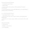
Indications for bioresorbable vascular scaffolds.
| Consensual and appropriate indications |
| 1. Young patients (aged <50 years) |
| 2. Diabetic patients |
| 3. Lesions in segments that may undergo CABG, particularly the left anterior descending artery |
| 4. Vessels with long lesions (>30 mm) and/or diffuse disease with a high probability of requiring secondary revascularization |
| Less consensual but possible indications |
| 1. Small collateral branches (<1.5 mm) |
| 2. Non-ST elevation acute coronary syndrome, stabilized, with intermediate and/or unstable plaques |
| 3. ST-elevation acute coronary syndrome, stabilized, with intermediate and/or unstable plaques |
| Inappropriate indications |
| 1. Left main disease |
| 2. Moderate or severe tortuosity |
| 3. Severe calcification |
CABG: coronary artery bypass grafting.
These guidelines are necessarily limited by constant developments in the state of the art and should be the subject of early review as technological advances and further evidence become available, preferably by a medical society specializing in the area.
Monitoring, research and costs in PortugalMedical societies in Portugal should organize and/or support research that includes multicenter registries and well-designed clinical trials, based on appropriate imaging and/or functional studies. There is no published information on the treatment's economic aspects, so assessment of the technological implications and incremental costs will be particularly relevant.
ConclusionThere is encouraging evidence that BVS are a viable treatment option. They should be used in more consensual indications and will require technical adaptations. Outcome monitoring and evaluation is essential to avoid inappropriate use. It is recommended that medical societies produce clinical guidelines as soon as possible.
Please cite this article as: Campante Teles R, Pereira H, Cyrne de Carvalho H, et al. Posição sobre suportes vasculares restaurativos transitórios coronários em Portugal. Rev Port Cardiol. 2013;32:1013–1018.
