


The prognostic value of late gadolinium enhancement (LGE) for risk stratification of hypertrophic cardiomyopathy (HCM) patients is the subject of disagreement. We set out to examine the association between clinical and morphological variables, risk factors for sudden cardiac death and LGE in HCM patients.
MethodsFrom a population of 78 patients with HCM, we studied 53 who underwent cardiac magnetic resonance. They were divided into two groups according to the presence or absence of LGE. Ventricular arrhythmias and morbidity and mortality during follow-up were analyzed.
ResultsPatients with LGE were younger at the time of diagnosis (p=0.046) and more often had a family history of sudden death (p=0.008) and known coronary artery disease (p=0.086). On echocardiography they had greater maximum wall thickness (p=0.007) and left atrial area (p=0.037) and volume (p=0.035), and more often presented a restrictive pattern of diastolic dysfunction (p=0.011) with a higher E/E′ ratio (p=0.003) and left ventricular systolic dysfunction (p=0.038). Cardiac magnetic resonance supported the association between LGE and previous echocardiographic findings: greater left atrial area (p=0.029) and maximum wall thickness (p<0.001) and lower left ventricular ejection fraction (p=0.056). Patients with LGE more often had an implantable cardioverter-defibrillator (ICD) (p=0.015). At follow-up, no differences were found in the frequency of ventricular arrhythmias, appropriate ICD therapies or mortality.
ConclusionsThe presence of LGE emerges as a risk marker, associated with the classical predictors of sudden cardiac death in this population. However, larger studies are required to confirm its independent association with clinical events.
O valor prognóstico do realce tardio na estratificação dos doentes com miocardiopatia hipertrófica é controverso. Este trabalho pretende avaliar a associação entre a presença de realce tardio na ressonância magnética cardíaca e características clínicas, imagiológicas e prognósticas em doentes com miocardiopatia hipertrófica.
MétodosDe 78 doentes com miocardiopatia hipertrófica avaliámos retrospetivamente 53, que realizaram ressonância cardíaca. Os doentes foram divididos em dois grupos, conforme a presença ou ausência de realce tardio. Foi feito seguimento clínico referente a disritmia ventricular e a morbi-mortalidade.
ResultadosOs doentes com realce tardio eram mais jovens à data do diagnóstico (p=0,046), mais frequentemente tinham antecedentes familiares de morte súbita (p=0,008) e de doença coronária (p=0,086). No ecocardiograma apresentavam maior espessura parietal máxima (p=0,007); área (p=0,037) e volume indexado da aurícula esquerda (p=0,035); maior frequência de padrão restritivo de disfunção diastólica (p=0,011), com relação E/E’ mais elevada (p=0,003); e disfunção sistólica do ventrículo esquerdo (p=0,038). A ressonância validou as alterações ecocardiográficas associadas à presença de realce tardio: maior área da aurícula esquerda (p=0,029); espessura parietal máxima (p<0,001) e menor fração de ejeção do ventrículo esquerdo (p=0,056). Os doentes com realce tardio mais frequentemente eram portadores de CDI (p=0,015); não havendo diferenças na frequência de episódios de disritmia ventricular, terapias apropriadas de CDI ou mortalidade no seguimento clínico.
ConclusõesA presença de realce tardio surge como um marcador de risco, associando-se a fatores já reconhecidos como preditores de morte súbita nesta população. A sua associação independente a eventos clínicos exige o estudo de populações de maior dimensão.
atrioventricular block
calcium channel blockers
cardiac magnetic resonance
hypertrophic cardiomyopathy
hazard ratio
implantable cardioverter-defibrillator
left atrial
late gadolinium enhancement
left ventricular
left ventricular ejection fraction
left ventricular hypertrophy
odds ratio
New York Heart Association
Hypertrophic cardiomyopathy (HCM) is a relatively common hereditary disease with a prevalence of 1/500 population, characterized by complexity and a wide spectrum of phenotypic expression and natural history.1,2
Sudden death is the leading cause of mortality in young HCM patients, although the risk is low (about 1%/year); an implantable cardioverter-defibrillator (ICD) is the only effective preventive measure.3 Risk stratification algorithms for predicting sudden death in this population have low positive predictive value and are unclear in their definition of risk factors.3,4
Cardiac magnetic resonance (CMR) has become an essential exam in the morphological and functional assessment of HCM. However, the prognostic value of late gadolinium enhancement (LGE) in identifying potentially arrhythmogenic areas of endomyocardial fibrosis is the subject of disagreement.5
The aim of this study was to examine the association between LGE on CMR and clinical, imaging and prognostic characteristics in a Portuguese population of HCM patients.
MethodsStudy populationWe performed a retrospective analysis of HCM patients followed regularly as outpatients in the cardiology department of a central hospital who underwent CMR with LGE study.
Of a population of 78 patients with HCM, 61 underwent CMR. The reasons for not undergoing CMR were the usual ones of ICD or pacemaker, claustrophobia, or patient refusal.
The definition of HCM was based on the classic echocardiographic criteria (left ventricular hypertrophy [LVH] without dilatation, maximum wall thickness ≥15 mm) and exclusion of other systemic or local causes of hypertrophy.
Eight patients were excluded: five in whom LGE study was not possible, one with a diagnosis of cardiac amyloidosis and two with Noonan syndrome. The final study population was thus composed of 53 patients.
Clinical dataThe patients’ clinical data were collected from their medical records by an investigator blinded to the results of CMR.
Baseline characteristicsThe following baseline characteristics were recorded: hypertension, diabetes, obstructive sleep apnea, coronary artery disease, atrial fibrillation, syncope, history of sudden death in a first-degree relative aged <45 years, New York Heart Association (NYHA) functional class, medication with beta-blockers or non-dihydropyridine calcium channel blockers, previous myectomy or alcohol septal ablation, pacemaker or ICD, resting electrocardiogram and NT-pro-BNP level.
Screening for Fabry disease was performed in 34 (64.1%) of the patients and for common HCM mutations in sarcomere protein genes in 41 (77.4%).
Assessment of blood pressure response by exercise testingThirty (56.6%) patients underwent exercise testing using the modified Bruce protocol. A hypotensive response was defined as a rise of ≤20 mmHg or a fall of ≥20 mmHg in blood pressure during exertion.
Echocardiographic parametersTransthoracic echocardiography was performed by two experienced observers who were blinded to some of the patients’ clinical data. The following parameters were assessed: left ventricular (LV) diastolic diameter; thickness of the ventricular septum, LV posterior wall and apex; left atrial (LA) volume; presence of a ≥30 mmHg gradient at rest and following provocation; quantification of mitral regurgitation; classification of LV systolic function and diastolic dysfunction.
Of the patients with no gradient at rest, 13 (33.4%) underwent exercise echocardiography to determine the presence of a gradient with exercise.
Cardiac magnetic resonanceAll CMR studies were performed on a Philips® 1.5 T scanner by three experienced observers who were blinded to some of the patients’ clinical data. The following parameters were assessed: LA area; left ventricular ejection fraction (LVEF); maximum LV wall thickness; and LGE following intravenous gadolinium administration.
Follow-upThe study population were followed for a mean of 53.6±53.4 months (4-271).
All patients underwent 24-hour Holter ECG monitoring to determine the number of ventricular extrasystoles and of episodes of non-sustained ventricular tachycardia, defined as ≥3 consecutive ventricular complexes lasting <30 s and without hemodynamic compromise.
In patients with an ICD, all records of device interrogation were analyzed for appropriate therapies and overdrive pacing triggered by ventricular fibrillation and/or tachycardia.
Two endpoints were defined: all-cause death and appropriate ICD therapies.
Statistical analysisThe statistical analysis was performed with SPSS for Windows, version 17.0.
Nominal variables were expressed as counts and percentages and were compared (combination of frequencies) using the chi-square test. Continuous variables were expressed as means ± standard deviation; the Student's t test was used to compare variables with normal distribution and the Mann–Whitney U test to compare those with non-normal distribution.
A value of p<0.05 was considered statistically significant.
ResultsCharacteristics of the study populationThe study population consisted of 53 patients, 27 (50.9%) male, with a mean age of 56.4±17.0 years at the time of diagnosis.
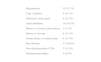
Table 1 shows the main characteristics of the population. In terms of functional capacity, 29 (54.7%) patients were in NYHA class I, 22 (41.5%) in class II, and two (3.8%) in class III. No patient had undergone myectomy and two had undergone alcohol septal ablation.
Main characteristics of the study population.
| Hypertension | 38 (71.7%) |
| Type 2 diabetes | 6 (11.3%) |
| Obstructive sleep apnea | 6 (11.3%) |
| Atrial fibrillation | 16 (30.2%) |
| History of coronary artery disease | 3 (5.7%) |
| History of syncope | 8 (15.1%) |
| Family history of sudden death | 9 (17.3%) |
| Beta-blockers | 37 (69.8%) |
| Non-dihydropyridine CCBs | 7 (13.2%) |
| Permanent pacemaker | 5 (9.4%) |
CCBs: calcium channel blockers.
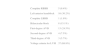
Table 2 shows the main electrocardiographic alterations.
Main electrocardiographic alterations.
| Complete RBBB | 5 (9.4%) |
| Left anterior hemiblock | 16 (30.2%) |
| Complete LBBB | 1 (1.9%) |
| Bifascicular block | 8 (15.1%) |
| First-degree AVB | 13 (24.5%) |
| Second-degree AVB | 4 (7.5%) |
| Third-degree AVB | 3 (5.7%) |
| Voltage criteria for LVH | 37 (69.8%) |
AVB: atrioventricular block; LBBB: left bundle branch block; LVH: left ventricular hypertrophy; RBBB: right bundle branch block.
Of the 34 (64.1%) patients screened for Fabry disease, none had any of the classical mutations.
Investigation of HCM mutations in sarcomere protein genes, performed in 41 patients (77.4%), was negative in 12 (29.3%); a classical mutation was identified in nine (21.9%), a mutation of undetermined significance in three (7.3%), and the results are still awaited in 17 (41.5%).
Mean NT-pro-BNP, assessed in 29 patients (54.7%), was 3130±5762 pg/ml.
Only four (13.3%) of the patients who underwent exercise testing had a hypotensive response.
Echocardiographic findingsThe phenotypic distribution of HCM was as follows: asymmetric in 36 (67.9%) patients, apical in 13 (24.5%) and concentric in four (7.5%). Systolic anterior motion of the mitral valve causing obstruction at rest was observed in 20 (37.7%) patients, with an end-systolic gradient of 69.4±27.3 mmHg. Mitral regurgitation was absent in six (11.3%) patients, minimal (grade 1) in 30 (56.6%), mild (grade 2) in 11 (20.8%), moderate (grade 3) in five (9.4%) and severe (grade 4) in one (1.9%).
Only seven (13.2%) patients did not present diastolic dysfunction. A pseudonormal pattern was the most common, found in 22 (41.5%) patients, followed by abnormal relaxation in 21 (39.6%); three (5.7%) presented a restrictive pattern.
At least mild global systolic dysfunction was identified in seven (13.2%) patients.
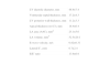
Table 3 shows the main continuous echocardiographic variables.
Main continuous echocardiographic variables.
| LV diastolic diameter, mm | 48.9±7.4 |
| Ventricular septal thickness, mm | 17.2±4.3 |
| LV posterior wall thickness, mm | 11.2±3.3 |
| Apical thickness (n=13), mm | 18.4±6.4 |
| LA area (A4C), mm2 | 25.1±5.6 |
| LA volume, mm3 | 52.5±20.1 |
| E-wave velocity, m/s | 0.82±0.31 |
| Lateral E′, cm/s | 6.7±2.4 |
| E/E′ ratio | 13.9±6.9 |
A4C: apical 4-chamber view; LA: left atrial; LV: left ventricular.
Of the 13 patients who underwent exercise echocardiography, seven (53.8%) developed a significant gradient on exertion (69.3±31.9 mmHg).
Cardiac magnetic resonance findingsThe phenotypic characterization of HCM by CMR was similar to that obtained by transthoracic echocardiography, and provided more accurate localization of LVH.
Table 4 shows the main continuous variables assessed by CMR.
LGE was observed in 24 (45.3%) patients.
Factors predicting late gadolinium enhancement on cardiac magnetic resonancePatients with LGE were younger at diagnosis (52.3±16.9 vs. 59.8±16.5 years; p=0.046), and more often had a family history of sudden death (33.3% vs. 3.4%; p=0.008; odds ratio [OR] 13.5).
Patients with LGE were more likely to have a history of coronary artery disease (12.5% vs. 0%; p=0.086) and higher NT-pro-BNP levels (5151±7882 vs. 1489±2422 pg/ml; p=0.089).
There were no differences between the groups in gender (p=0.669); history of hypertension (p=0.899), diabetes (p=0.532), syncope (p=0.288) or obstructive sleep apnea (p=0.135); presence of an identified mutation (p=0.676); NYHA functional class; previous therapy with beta-blockers (p=0.454) or non-dihydropyridine CCBs (p=0.112); voltage criteria for LVH (p=0.696); or history of atrial fibrillation (p=0.098).
There was also no difference between the groups in blood pressure response to exercise testing (p=0.348).
Patients with LGE more often presented systolic dysfunction on echocardiography, characterized by LVEF <50% (25.0% vs. 3.4%; p=0.038; OR 9.33) and diastolic dysfunction with pseudonormal (68.2% vs. 29.2%) or restrictive pattern (9.1% vs. 4.2%; p=0.011).
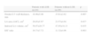
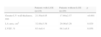
Table 5 shows the continuous echocardiographic variables that predicted LGE.
Continuous echocardiographic variables that predicted late gadolinium enhancement.
| Patients with LGE (n=24) | Patients without LGE (n=29) | p | |
| Greater LV wall thickness, mm | 18.96±5.08 | 15.61±2.82 | 0.007 |
| LA area (A4C), cm2 | 26.85±5.97 | 23.57±4.94 | 0.037 |
| Indexed LA volume, cm3 | 58.97±20.77 | 47.05±18.13 | 0.035 |
| E/E′ ratio | 16.73±7.72 | 11.32±4.96 | 0.003 |
A4C: apical 4-chamber view; LA: left atrial; LGE: late gadolinium enhancement.
No association was seen between the presence of LGE and type of HCM: asymmetrical (p=0.441), apical (p=0.475) and concentric (p=0.844). Similarly, neither the presence of obstruction (p=0.242) nor the severity of mitral regurgitation (p=0.637) was associated with LGE.
Table 6 shows the CMR variables that predicted LGE.
Cardiac magnetic resonance variables that predicted late gadolinium enhancement.
| Patients with LGE (n=24) | Patients without LGE (n=29) | p | |
| Greater LV wall thickness, mm | 21.50±4.05 | 17.69±2.57 | <0.001 |
| LA area, cm2 | 32.88±3.58 | 29.00±5.26 | 0.029 |
| LVEF, % | 63.4±8.4 | 68.1±8.8 | 0.056 |
LA: left atrial; LGE: late gadolinium enhancement; LVEF: left ventricular ejection fraction.
Patients were followed for a mean of 53.6±53.4 months (4-271).
There were no differences between the groups in detection of non-sustained ventricular tachycardia (20.8% vs. 20.7%; p=0.990), or in the number of ventricular extrasystoles (p=0.503) documented on 24-hour Holter monitoring.
During follow-up 11 patients (20.7%) received an ICD for primary prevention due to the presence of one or more classical risk factors. More patients with LGE received ICDs (37.5% vs. 6.9%; p=0.015). An appropriate therapy with overdrive pacing was recorded in only one patient.
Two patients died during follow-up, one in each group, both from heart failure.
DiscussionCMR is the preferred imaging modality for determining ventricular mass, chamber volume, global systolic function and the pattern and distribution of LVH in HCM patients.6–8 LGE imaging is a particularly valuable method for identifying areas of fibrosis and to determine their extent and distribution.9,10
Characteristics of the study populationThe mean age of our population (56.4±17.0 years) was older than in most studies in the literature5,11,12 and included few patients in the age-group most associated with sudden death (<35 years).
Most were asymptomatic or mildly symptomatic, as in other published series.9,13–15 This highlights the need for new ways to stratify risk of sudden death, since this may be the first clinical manifestation of HCM, years after initial diagnosis.16,17
Although asymmetric HCM was the most common pattern in our population (67.9%), there was a high prevalence of apical HCM (24.5%).
The mean maximum LV wall thickness on CMR of 19.4±3.8 mm demonstrates that significant LVH can be present even in only mildly symptomatic patients, as reported in the literature.13–15
The prevalence of LV outflow tract obstruction in our population (50.9%) was lower than in the literature,18 but some patients without a gradient at rest did not undergo exercise echocardiography.
The percentage of patients with a hypotensive response on exercise testing (13.3%) was also lower than previously reported (25%).19,20
Clinical and prognostic significance of late gadolinium enhancementOur study confirms that the presence of LGE is a common phenotypic characteristic in this population, although its prevalence (45.3%) was lower than that described in the literature.21–23
The pattern of distribution of LGE does not correspond to the perfusion territories of the epicardial coronary arteries, but is related to areas of LVH and is mid-myocardial in location.9
Predictors of LGE were younger age, a family history of sudden death, LVEF <50%, more severe diastolic dysfunction and LA dilatation, and greater maximum LV wall thickness.
It is known that the presence and extent of LGE is inversely related to LVEF,24 since LGE is associated with ventricular remodeling that eventually leads to heart failure.13 An association between LGE and maximum LV wall thickness has also been reported.25 LA dilatation, a surrogate marker of diastolic dysfunction, is also associated with LGE.26
Mortality in our population, although low, was in all cases of cardiovascular cause (heart failure).
As in previous studies,11,15 it remains to be proved whether LGE is an independent prognostic marker. Maron et al.12 reported a high prevalence, although not reaching statistical significance, of cardiovascular events during follow-up of 202 patients with HCM and LGE. By contrast, in a study of 220 asymptomatic or only mildly symptomatic HCM patients with a mean follow-up of three years, Bruder et al.15 showed that LGE was an independent predictor of cardiac death (hazard ratio [HR] 8.6; p=0.038), whereas the presence of one or two clinical risk factors did not reach statistical significance (HR 1.4; p=0.68).
In a recent meta-analysis of four studies with a total of 1063 HCM patients, Green et al.5 confirmed the high prevalence of LGE (60%) and its correlation with adverse cardiovascular events (cardiac death, heart failure death, and all-cause mortality), although it was not associated with sudden death or aborted sudden death and did not add value to traditional clinical risk factors.
In a population in which 60% present LGE and the event rate is 1–5%/year, such an association would be difficult to prove. More sophisticated techniques for quantifying LGE would thus improve its prognostic value.27 Quantification of the extent of LGE by means of T1-weighted sequences has been validated in other diseases of the myocardium, including ischemic heart disease and dilated cardiomyopathy, and could be of value in HCM.28,29
The European Cardiovascular Magnetic Resonance registry, with a clinical follow-up of at least 12 months, observed only a trend towards better outcome in HCM patients without LGE.30
Study limitationsOur study has several limitations, including the small number of patients and events.
Patients with ICDs who did not undergo CMR before device implantation were not included in the study, and it is impossible to know whether their inclusion would have significantly changed its results.
Another limitation is the fact that LGE was not quantified, nor was its distribution analyzed.
As with all retrospective studies, we were limited to the information available in patients’ medical records.
ConclusionsThe presence of LGE emerges as a risk marker, associated with the classical predictors of sudden cardiac death in HCM, particularly a family history of sudden death and greater maximum LV wall thickness. However, larger studies are required to confirm its independent association with clinical events.
Ethical disclosuresProtection of human and animal subjectsThe authors declare that no experiments were performed on humans or animals for this study.
Confidentiality of dataThe authors declare that they have followed the protocols of their work center on the publication of patient data.
Right to privacy and informed consentThe authors have obtained the written informed consent of the patients or subjects mentioned in the article. The corresponding author is in possession of this document.
Conflicts of interestThe authors have no conflicts of interest to declare.
Please cite this article as: Caetano F, Botelho A, Trigo J, et al. Expressão fenotípica da miocardiopatia hipertrófica e realce tardio na ressonância magnética cardíaca. Rev Port Cardiol. 2014;33:261–266.
