


A 67-year-old man, who underwent a right nephrectomy seven months ago due to renal cell carcinoma, presented with fever, Staphylococcus hominis bacteriemia and decompensated congestive heart failure.
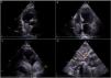
An abdominal computed tomography (CT) revealed a distended inferior vena cava, which was assumed to be probable venous thrombosis. To exclude endocarditis, a transthoracic echocardiography (TEE) was performed, showing reduced ejection fraction (38%) due to global hypokinesia, and a large heterogeneous mass occupying almost the entire right atrium (Figure 1). The mass did not cause obstruction of the tricuspid valve but extended to the inferior vena cava. There were no signs of endocarditis.

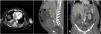
The patient was then diagnosed with inferior vena cava syndrome due to vascular metastasis with associated thrombus and was transferred to the intensive care unit. Following 72 hours of unfractionated heparin perfusion, the repeat CT revealed no changes in the mass extension (Figure 2). After discussion with the vascular surgeon, cardiothoracic surgeon, urologist and oncologist, the patient is under evaluation for surgical resection of the tumor.

Renal cell carcinoma accounts for 3% of global cancer diagnosis, and its incidence has been increasing in the past years.1 Although isolated cardiac metastasis is rare, the tumor can extend up the inferior vena cava, presenting typically as a “fingerlike” projection protruding into the right atrium on echocardiography.2 Following its origin with echocardiography into the inferior vena cava may to help differentiate it from other cardiac masses.
This case highlights the importance of carefully considering the whole clinical context when evaluating a cardiac mass.
Conflicts of interestThe authors have no conflicts of interest to declare.
