Suggestions
Idioma
Journal Information
Vol. 44. Issue 9.
Pages 591-593 (September 2025)
Vol. 44. Issue 9.
Pages 591-593 (September 2025)
Image in Cardiology
Full text access

Mitral valve aneurysm, an unusual lesion not to miss

Aneurisma da válvula mitral, uma lesão incomum a não perder
Visits
1713
Cristhian Emilio Herreraa,
Corresponding author
emilio.herrera@udea.edu.co

Corresponding author.
, Jhon F. Salamancab, Luisa F. Durangob
a Clinical Cardiology Department, Cardiovascular Unit, Clínica Medellín, Medellín, Colombia
b Echocardiography Department, Cardiovascular Imaging Unit, Clínica CardioVID, Medellín, Colombia
This item has received
Article information
Full Text
Bibliography
Download PDF
Statistics
Figures (2)
fig0005
fig0010
Additional material (4)
Full Text

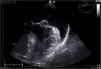
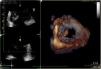
We present the case of a 65-year-old man with a history of high blood pressure. During a follow-up appointment, a holosystolic murmur, III/VI, radiating to the armpit was detected. He was cardiovascularly asymptomatic. A complete echocardiogram was performed, with the following findings: severely dilated left atrium; mitral valve with an aneurysmal lesion at the A2 scallop (Figure 1) causing severe regurgitation with an eccentric jet. There was systolic flow reversal in the pulmonary veins. The mechanism of regurgitation is Carpentier II. The rest of the valves had normal morphology and function, biventricular function, large vessels, and the pericardium. There was an intermediate probability of pulmonary hypertension. Findings were confirmed and the lesion was further characterized using transesophageal echocardiogram and three-dimensional reconstruction (Figure 2).

Figure 1.

Transesophageal echocardiogram, mid-esophageal view at 90°, showing a mitral valve aneurysm with dimensions of 1.5 cm and 1.2 cm, causing a defect and an 8 mm displacement over the annular plane.

Figure 2.

Three-dimensional reconstruction, transesophageal echo, and mitral valve view from the left atrium, depicting a saccular aneurysmal lesion on the A2 scallop.

The patient opted not to undergo surgery. He is under clinical and echocardiographic follow-up every three months and remains asymptomatic.

Mitral valve aneurysm is a rare condition, with a prevalence estimated between 0.02 and 0.29%.1 It can cause valve regurgitation and ventricular dysfunction with multiple consequences. It is described as a saccular lesion with thin walls protruding into the atrium during systole. Its origin has been mainly associated with endocarditis, primarily of the aortic valve, with worse prognostic implications. However, an infectious origin has not been found in all cases.2

It is important to consider mitral valve aneurysm in the differential diagnosis of mitral lesions (endocarditis, tumors, thrombi, among others).3

Conflicts of interest

The authors have no conflicts of interest to declare.

Appendix B
Supplementary data

The following are the supplementary data to this article:

(0.1MB)
(0.45MB)
(0.3MB)
(0.15MB)

References
[1]
J.F. Vasquez-Rodriguez, A. Martínez-Caballero, O.M. Perez-Fernandez, et al.
Unusual lesions of the mitral valve: two different conditions with the same imaging findings.
CASE, 3 (2019), pp. 204-209
[2]
I. Vilacosta, J.A. San Román, C. Sarriá, et al.
Clinical, anatomic, and echocardiographic characteristics of aneurysms of the mitral valve.
Am J Cardiol, 84 (1999), pp. 110-113
[3]
M.M. Stechert, J.R. Pletcher, E.E. Tseng, et al.
Aneurysm of the anterior mitral valve.
Anesth Analg, 114 (2012), pp. 86-88
Copyright © 2025. Sociedade Portuguesa de Cardiologia
Download PDF
Idiomas
Revista Portuguesa de Cardiologia (English edition)
Article options
Tools
Supplemental materials