Journal Information
Vol. 43. Issue 3.
Pages 147-148 (March 2024)
Vol. 43. Issue 3.
Pages 147-148 (March 2024)
Image in Cardiology
Full text access
Lung adenocarcinoma: The diagnostic importance of echocardiography
Adenocarcinoma do pulmão: importância diagnóstica da ecocardiografia
Visits
2702
Inês Ferreira Neves1,
Corresponding author
inessferreiraneves@gmail.com

Corresponding author.
, Pedro Garcia Brás1, Pedro Coelho, Paulo Calvinho, Luísa Branco
Department of Cardiology, Hospital de Santa Marta, Centro Hospitalar Universitário de Lisboa Central, Lisbon, Portugal
This item has received
Article information
Full Text
Bibliography
Download PDF
Statistics
Figures (1)
Additional material (2)
Full Text

A 44-year-old man with poorly differentiated lung adenocarcinoma (stage IV, T4N0M1a) was referred for a transthoracic echocardiogram (TTE) to assess for cardiac metastasis. Although there were no signs of left atrial invasion in parasternal and apical views, subcostal view on the TTE showed an image suggestive of a cardiac mass, with protrusion to the left atrium (LA) through the right inferior pulmonary vein (RIPV) (Figure 1A and Video 1). There were no other abnormal findings on the TTE. To clarify the extension of the cardiac mass, a transesophageal echocardiogram (TEE) was performed, revealing an exuberant tumoral mass in the RIPV, measuring 8.2 cm, with protrusion of 2.8 cm into the LA (Figure 1B and Video 2). Despite an apparent surgical cleavage plane in the LA, the cardiac mass showed complete invasion of the RIPV wall (Figure 1B). There were no masses adjacent to the left pulmonary veins. A chest computed tomography (CT) scan confirmed tumor invasion through the RIPV (Figure 1C and D). The patient then underwent successful surgical tumorectomy of the lung and RIPV mass and atrioplasty. The postoperative course was uneventful.

Figure 1.

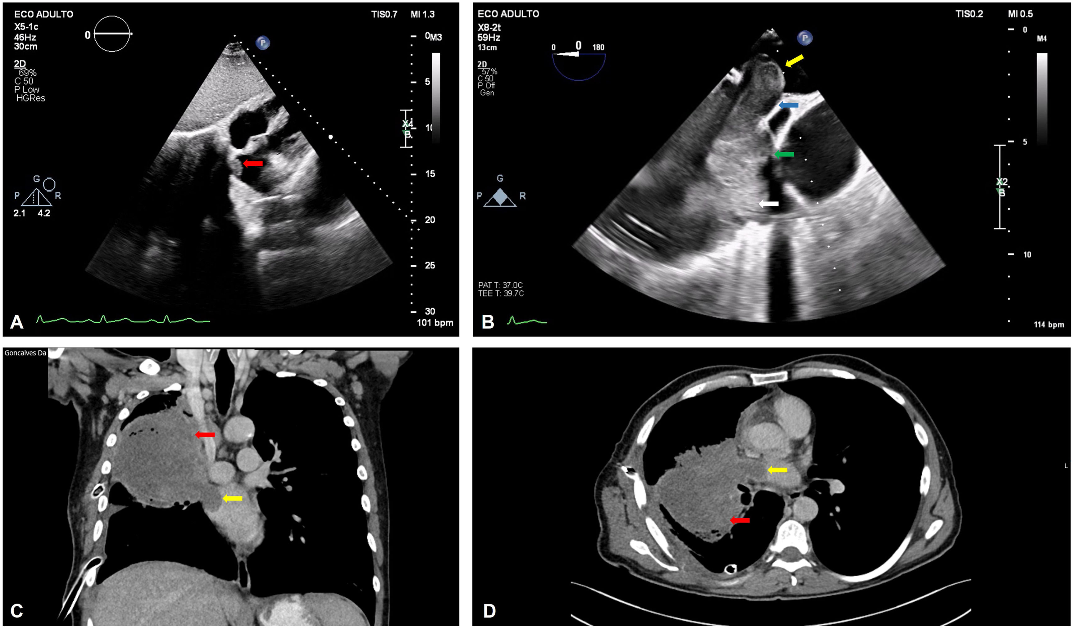
(A) Transthoracic echocardiogram (TTE) with an image suggestive of a cardiac mass with protrusion to the left atrium (LA) (red arrow); (B) TEE with an exuberant tumoral mass in the right inferior pulmonary vein (RIPV) with protrusion of 2.8 cm into the LA (yellow arrow); apparent surgical cleavage plane in the LA (blue arrow); area without surgical cleavage plane (green arrow); lung carcinoma (white arrow); (C) (coronal plane) and (D) (axial plane): chest computed tomography scan showing lung carcinoma (red arrow) invading the LA (yellow arrow).

Cardiac metastasis from lung carcinomas is rare.1 TTE is a crucial initial imaging modality as it can detect cardiac invasion to the pericardium or through the pulmonary veins.2,3 However, a high level of suspicion is needed, as a thorough scan may uncover minor findings that should prompt further imaging. CT, cardiac magnetic resonance imaging and positron emission tomography can provide further information on differential diagnosis of the mass.2–4

Conflicts of interest

The authors have no conflicts of interest to declare.

Appendix A
Supplementary data

The following are the supplementary data to this article:

(0.47MB)
(0.19MB)

References
[1]
I. Burazor, S. Aviel-Ronen, M. Imazio, et al.
Metastatic cardiac tumors: from clinical presentation through diagnosis to treatment.
BMC Cancer, 18 (2018), pp. 202
[2]
D. Auger, J. Pressacco, F. Marcotte, et al.
Cardiac masses: an integrative approach using echocardiography and other imaging modalities.
Heart, 97 (2011), pp. 1101-1109
[3]
O. Buckley, R. Madan, R. Kwong, et al.
Cardiac masses, part 1: imaging strategies and technical considerations.
AJR Am J Roentgenol, 197 (2011), pp. W837-W841
[4]
A.D. Goldberg, R. Blankstein, R.F. Padera.
Tumors metastatic to the heart.
Circulation, 128 (2013), pp. 1790-1794

These authors contributed equally to this work.

Copyright © 2023. Sociedade Portuguesa de Cardiologia
Download PDF
Idiomas
Revista Portuguesa de Cardiologia (English edition)
Article options
Tools
Supplemental materials
en pt

Are you a health professional able to prescribe or dispense drugs?

Você é um profissional de saúde habilitado a prescrever ou dispensar medicamentos

By checking that you are a health professional, you are stating that you are aware and accept that the Portuguese Journal of Cardiology (RPC) is the Data Controller that processes the personal information of users of its website, with its registered office at Campo Grande, n.º 28, 13.º, 1700-093 Lisbon, telephone 217 970 685 and 217 817 630, fax 217 931 095, and email revista@spc.pt. I declare for all purposes that the information provided herein is accurate and correct.