


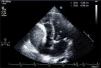
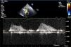

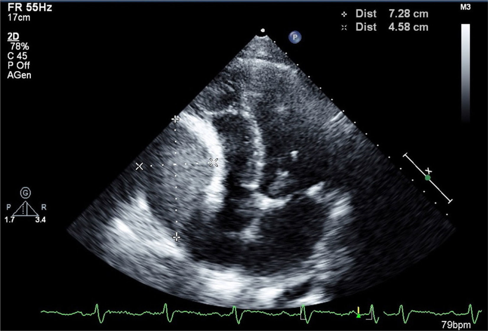
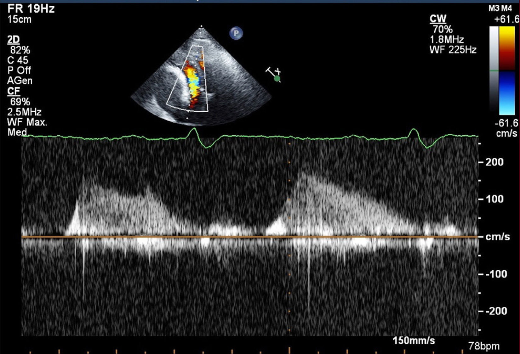
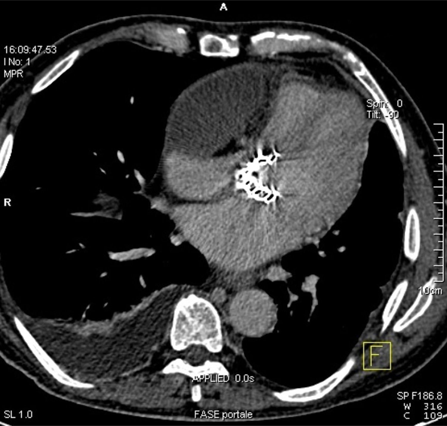
An 82-year-old Caucasian man presented with a history of worsening exertional dyspnea. Two years before the referral he had undergone transapical aortic valve implantation due to severe aortic stenosis. Transthoracic echocardiography was promptly performed: it demonstrated a mild pericardial effusion and a 7.3×4.6 cm homogeneous mass causing extrinsic compression of the right atrium (RA) (Figure 1) and severe functional tricuspid valve stenosis (mean gradient 6 mmHg) (Figure 2). To better define the mass and its relationship with adjacent cardiac structures, contrast chest computed tomography was performed, which confirmed mild pericardial effusion without pericardial thickening and revealed a low-density encapsulated mass arising from the wall of the RA and extending across the right atrioventricular groove (Figure 3), without enhancement.
At thoracotomy, the parietal pericardium appeared normal, and 200 ml of straw-colored fluid was recovered from the pericardial cavity. The fluid was negative for malignant cells and consisted of a few benign and reactive mesothelial cells mixed with inflammatory cells. A cystic mass was located anterior and inferior to the RA, extending to the right ventricle and covered by thickened fibrous tissue, pericardium and epicardium. The right atrial and ventricular surface was covered with thick, white, glistening epicardium.
The mass was removed and a waffle procedure (multiple longitudinal and transverse incisions in the thickened epicardium) was performed without cardiopulmonary bypass.
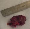
The biopsy, performed after surgical removal (Figure 4), revealed lymphoplasmacytic infiltrate diagnostic of epicarditis.

The patient's postoperative course was uneventful and his symptoms improved markedly.
Ethical disclosuresProtection of human and animal subjectsThe authors declare that no experiments were performed on humans or animals for this study.
Confidentiality of dataThe authors declare that no patient data appear in this article.
Right to privacy and informed consentThe authors declare that no patient data appear in this article.
Conflicts of interestThe authors have no conflicts of interest to declare.
