Suggestions
Idioma
Journal Information
Vol. 44. Issue 11.
Pages 703-704 (November 2025)
Vol. 44. Issue 11.
Pages 703-704 (November 2025)
Image in Cardiology
Full text access

Left atrial appendage aneurysm: An unexpected finding in a patient with asymptomatic atrial fibrillation

Aneurisma do apêndice auricular esquerdo: um achado inesperado num doente com fibrilhação auricular assintomática
Visits
843
Javier Cantalapiedra Romeroa,b,
Corresponding author
jcantalapiedraromero@gmail.com

Corresponding author.
, Carlos Izurietaa,b, Marta Zielonkaa,b
a Hospital Universitario Arnau de Vilanova, Lleida, Spain
b Institut Recerca Biomèdica (IRB), Lleida, Spain
This item has received
Article information
Full Text
Bibliography
Download PDF
Statistics
Figures (2)
fig0005
fig0010
Additional material (2)
Full Text

Our case was a 48-year-old woman with no prior medical history referred due to asymptomatic atrial fibrillation (AF) of several years in duration. A baseline electrocardiogram showed a controlled heart rate without medication and no other abnormalities.

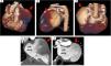
A transthoracic echocardiogram revealed a large saccular structure communicating with the left atrium, with Doppler flow inside it (Figure 1 and supplementary videos 1 and 2), but no other abnormalities were identified. Further evaluation with computed tomography confirmed the presence of a left atrial appendage aneurysm (8 cm×5 cm) with no thrombus inside and normal coronary arteries (Figure 2).

Figure 1.

4-Chamber apical view showing (A) left atrial appendage aneurysm (red arrow) with Doppler flow within it (B).

Figure 2.

Computed tomography showing the anatomic reconstruction of the left atrial appendage aneurysm (red arrow) from the following perspectives. (A) Anterior, (B) lateral, (C) posterior, (D) slow contrast flow within the aneurysm and (E) absence of thrombi inside it.

Electrical cardioversion was unsuccessful, leading to the abandonment of a rhythm-control strategy. In agreement with the patient and given the absence of symptoms or signs of compression of adjacent structures, a conservative management approach was chosen. Oral anticoagulation with dabigatran was prescribed due to the medium-to-long-term thromboembolic risk.

This case highlights the importance of ruling out structural abnormalities in young patients with persistent AF. Left atrial appendage aneurysm is a rare finding, predominantly reported as an asymptomatic condition in women in their 30s and 40s.1 It may be congenital, 90% due to pectinate muscle dysplasia, or acquired, resulting from herniation through a pericardial defect or increased atrial pressures due to mitral valve disease.2

The differential diagnosis includes entities such as coronary fistula or a pericardial cyst, and multimodal imaging is crucial for accurate management. Treatment for symptomatic cases is typically surgical; however, no studies are available comparing surgery versus conservative management with anticoagulation in asymptomatic patients.3

Conflicts of interest

The authors have no conflicts of interest to declare.

Appendix B
Supplementary data

The following are the supplementary data to this article:

(0.14MB)
(0.31MB)

References
[1]
Y. Daralammouri, A. Odeh, S. Abuzahra, et al.
Left atrial appendage aneurysm: a descriptive systematic review of 177 cases.
BMC Cardiovasc Disord, 24 (2024), pp. 633
[2]
J.D. Ayala Torres, J.A. Sepulveda Gallego, M. Gonzalez Gonzalez.
Left atrial appendage aneurysm: a case report and literature review.
Cureus, 16 (2024), pp. e56280
[3]
A. Mittal, M. Navaratnarajah, S. Harden, et al.
Staged hybrid ablation in left atrial appendage aneurysm a rare cause of refractory atrial tachyarrhythmia – a case report.
Eur Heart J Case Rep, 8 (2024), pp. 1-6
Copyright © 2025. Sociedade Portuguesa de Cardiologia
Download PDF
Idiomas
Revista Portuguesa de Cardiologia (English edition)
Article options
Tools
Supplemental materials