


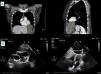
A 50-year-old woman, asymptomatic and with no relevant prior history, underwent an occupational medical examination. She was diagnosed with latent tuberculosis infection, so she was referred for a chest X-ray, which identified a mediastinal mass. She underwent a computed tomography scan in which a well-defined cystic lesion was discovered in the left costophrenic angle, about 8 cm×7 cm×10 cm in size (Figure 1A).

She was referred for a cardiology consultation to assess the hemodynamic compromise caused by the mass. A transthoracic echocardiogram (Figure 1B) confirmed the cystic lesion in the left costophrenic angle (9 cm×7 cm). It had well-defined edges and two septa inside with echolucent content, and caused mild extrinsic compression of the lateral and apical walls of the left ventricle without compromising filling of the cavity. Infiltration of the adjacent cavities was not observed, confirming the diagnosis of uncomplicated left pleuropericardial cyst.
This case report is of considerable interest due to the size and unusual location of the pleuropericardial cyst. Although this is an uncommon entity, with an incidence of 1/100000 individuals, it is undeniably relevant. Usually, pleuropericardial cyst is a benign condition and diagnosis is incidental. In our case, the location was atypical, being in the left costophrenic angle (20%), where it is closely related to the heart; such cases are most often associated with complications, mainly due to hemodynamic compromise. The differential diagnosis is broad, including other mediastinal masses. The rarity of this presentation and the possible complications add complexity to the management of this condition.
Conflicts of interestThe authors have no conflicts of interest to declare.
