

Myotonic dystrophy is a multisystem disease associated with cardiac abnormalities that are responsible for high morbidity and mortality. It commonly affects conduction tissue, resulting in changes in heart rate that tend to progress with age.
ObjectiveThe aim of the study was to assess overall cardiovascular risk and the risk of arrhythmias in patients with myotonic dystrophy type 1 (DM-1) and to correlate them with genetic study (CTG expansion size).
MethodsThis retrospective study included 31 DM-1 patients referred to the cardiology department of Centro Hospitalar Entre Douro e Vouga by the neurology department for screening for heart disease. Patients’ medical records were consulted for the diagnostic tests performed in the diagnostic cardiology consultation: electrocardiogram (ECG), high-resolution ECG, heart rate variability (HRV), Holter 24-hour ambulatory ECG and transthoracic echocardiogram (TTE); results of genetic testing were also consulted.
ResultsOf 31 patients studied, 38% had first-degree atrioventricular block (AVB) and 51% had intraventricular conduction disturbances (62% had late potentials). TTE revealed no structural heart disease. Rare supraventricular and ventricular ectopic beats were the most common arrhythmias on 24-hour Holter monitoring. The sample showed lower HRV, reflecting vagal dysfunction. Patients with larger CTG expansions had more cardiac abnormalities.
ConclusionsPatients with DM-1 had arrhythmic events, with AVB and more significantly intraventricular block, although none had malignant arrhythmias or structural heart disease. No patient died. Patients with larger CTG expansions had greater involvement of cardiac conduction tissue.
A distrofia miotónica é uma doença multissistémica, que se associa a alterações cardíacas que são responsáveis por elevada taxa de morbilidade e mortalidade. O atingimento do tecido de condução é frequente, traduzindo-se em alterações no ritmo cardíaco, que tendem a progredir com a idade.
ObjetivoO presente trabalho propõe-se avaliar o perfil cardiovascular global, bem como o risco de arritmias cardíacas, em indivíduos com distrofia miotónica de steinert tipo 1 (DMS1), correlacionando os diversos achados com o estudo genético (tamanho da expansão CTG) comparando possíveis diferenças entre géneros.
MétodosEstudo retrospetivo que incluiu 31 pacientes DMS1 referenciados ao serviço de cardiologia do Centro Hospitalar Entre Douro e Vouga pelo serviço de neurologia para rastreio de doença cardíaca. Todos os doentes realizaram uma consulta clínica e foram submetidos a exames complementares de diagnóstico, nomeadamente eletrocardiograma (ECG), eletrocardiograma de alta resolução (ECGAR), variabilidade da frequência cardíaca (VFC), monitorização eletrocardiográfica ambulatória contínua (MEAC-Holter) e ecocardiograma transtorácico (ETT). Foi ainda consultado o exame genético previamente efetuado.
ResultadosDos 31 doentes estudados, 38% apresentaram BAV 1.° grau e 51% apresentaram perturbação da condução intraventricular no ECG; 62% dos doentes apresentaram potenciais ventriculares tardios; não se verificou doença cardíaca no ETT; as extrassístoles supraventriculares e ventriculares raras foram as arritmias mais frequentes no Holter de 24 h; a amostra apresentou valores diminuídos para a VFC, o que reflete a presença de disfunção vagal. Os doentes com maiores expansões CTG apresentaram maior número de anormalidades cardíacas.
ConclusõesPacientes com DMS1 apresentam alterações cardíacas, embora a gravidade das alterações documentadas esteja aquém do sugerido na literatura. Este aspeto poderá refletir a qualidade do seguimento clínico oferecido aos doentes por um centro com uma consulta especializada.
Myotonic dystrophy type 1 (DM-1), also known as Steinert disease, is a genetic disease caused by abnormal expansion of the cytosine-thymine-guanine (CTG) triplet in the DMPK (dystrophia myotonica-protein kinase) gene on chromosome 19, which is mainly expressed in skeletal and cardiac muscle.1 DM-1 is the commonest form of muscular dystrophy in adults, with a prevalence of 2–14:100000 individuals and an incidence of 1:8000.2–5 It is a hereditary multisystemic disease with autosomal dominant transmission and incomplete penetrance.
Although the main clinical manifestations of DM-1 are myotony and muscle weakness secondary to involvement of skeletal muscle, other organs and systems may be affected, particularly the cardiovascular and respiratory systems.6 The reduced life expectancy of these patients is largely due to high mortality resulting from respiratory infections, cancer and sudden cardiac death (SCD).5
Heart disease is common in DM-1, although its actual prevalence is difficult to estimate. There is evidence that conduction disturbances are the most frequent cardiac manifestation, but other complications have been reported, including tachyarrhythmias.6,7 Some findings also indicate that progressive muscular impairment is associated with worsening arrhythmic events, the prevalence of which can reach 80% of cases.8 Arrhythmias are the second leading cause of death in DM-1 after pneumonia, causing up to 30% of deaths. Most patients (over 60%) who die from cardiac cause suffer SCD.9
In view of the impact of cardiac manifestations on the survival of DM-1 patients, it is important to identify potential predictors of SCD to enable appropriate cardiovascular risk stratification. Some authors argue that the degree of neurological involvement and CTG expansion size are directly related to the severity of conduction tissue disease.10–12 On the other hand, major alterations on the electrocardiogram (ECG) have been proposed as an independent predictor of SCD in these patients. However, the nature of conduction disturbances in DM-1, together with the fact that intermittent conduction and rhythm abnormalities are observed in a significant number (32%) of patients with normal ECG, mean that 24-hour Holter monitoring to identify and quantify arrhythmias is an important element in the management of these patients.5,13 Evidence for the usefulness of high-resolution ECG in this context is inconclusive; the technique has high sensitivity but low specificity for ventricular arrhythmias.3 Measures of cardiac autonomic modulation, particularly heart rate variability (HRV), have also been studied, but their predictive value for SCD in DM-1 has not been established.14 Transthoracic echocardiography (TTE) is also used to screen for heart disease in these patients, although cardiomyopathy is rare in DM-1 and studies to date have provided limited information.3
The aim of the study was to assess the risk of arrhythmias in patients with DM-1, to identify possible gender differences and to characterize overall cardiovascular risk, and to correlate the latter with genetic study (CTG expansion size).
MethodsWe performed a retrospective study (between 2000 and 2011) of 34 DM-1 patients diagnosed with DM-1 referred to the cardiology department of Centro Hospitalar Entre Douro e Vouga by the neurology department for screening for heart disease, who underwent ECG, high-resolution ECG, TTE, 24-hour Holter monitoring and HRV assessment. After application of the exclusion criteria (other types of myotony or DM or failure to complete the consultation protocols), the final study population consisted of 31 patients.
Standard 12-lead ECG was performed and the following information was recorded: heart rhythm; heart rate; QRS axis; and PR, QRS, QT, and QTc intervals. All the ECG recordings were assessed independently by two experienced observers in accordance with international guidelines.15
High-resolution ECG was also performed in the standard fashion,16 and the following variables were recorded: filtered QRS complex duration (QRSd), root mean square voltage of the last 40 ms of the QRS complex (RMS40), and duration of low-amplitude signals (<40 mV) (LAS40) in the terminal QRS. Ventricular late potentials (VLP) were considered present when two of the following parameters were considered abnormal compared to the reference values: QRSd <114 ms; RMS40 >20 μV and LAS40 <38 ms.17
TTE, performed in accordance with the guidelines,18 was used to assess the following variables: valve morphology and function; cardiac chamber dimensions; ventricular wall thickness; and global left ventricular (LV) systolic function as assessed by LV ejection fraction (LVEF), velocity of circumferential fiber shortening (VCF), a measure of myocardial contractility, and LV mass index (LVMI).
From 24-hour Holter monitoring, performed according to the guidelines,18 conduction and rhythm disturbances were identified, using Lown's classification to characterize ventricular ectopic beats. Analysis of HRV, based on the Holter recordings, included the following time-domain indices: standard deviation of NN intervals (SDNN), standard deviation of average 5-min NN intervals (SDANN), root mean square of successive differences of NN intervals (RMSSD), and the percentage of pairs of NN intervals differing by >50 ms (pNN50). Frequency-domain parameters assessed were high-frequency (HF) and low-frequency (LF) spectral power and LF/HF ratio.
For analysis of the correlation between CTG expansion size and cardiac events, the sample was divided into two groups, equal to or below and above the median of repeats in the sample (900).
Statistical analysisSPSS® version 19.0 was used to perform a descriptive analysis. Possible gender differences were investigated. Quantitative variables were presented as means ± standard deviation and coefficient of variation, and qualitative variables as absolute frequencies.
The means of quantitative variables for both sexes and correlation with CTG expansion size were compared using the Student's t test for independent samples, and the chi-square test or Fisher's exact test, as appropriate, were used for analysis of categorical variables.
A value of p<0.05 for a 95% confidence interval was adopted as the criterion of statistical significance.
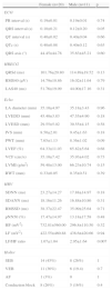
ResultsPopulation characteristicsThe final sample consisted of 31 patients, of whom 20 (64.5%) were female and 11 (35.5%) were male. Mean age was 42.0±13.1 years (range 18–66). Mean body mass index (BMI) was 27.14±4.72 kg/m2 and was higher in women than in men (29.36±5.11 kg/m2 vs. 25.75±4.08 kg/m2; p=0.04), while mean weight, height and body surface area were higher in men (Table 1).
Anthropometric characteristics of the study population (n=31) by gender.
| Women (n=20) | Men (n=11) | p | |
| Age (years) | 43.35±13.42 | 39.55±12.7 | 0.44 |
| Weight (kg) | 70.60±11.94 | 71.00±10.32 | 0.92 |
| Height (cm) | 155.20±3.69 | 166.27±4.94 | <0.001 |
| BSA (m2) | 1.69±0.12 | 1.78±0.11 | 0.05 |
| BMI (kg/m2) | 29.36±5.11 | 25.75±4.08 | 0.04 |
BMI: body mass index; BSA: body surface area.
Nineteen patients (61%) presented normal ECG. Of the others, 12 (38%) had first-degree atrioventricular block (AVB), nine (29%) of them women and three (9.7%) men. Intraventricular conduction disorders were found in 16 patients (51%), nine (29%) of them women and seven (23%) men. Mean QRS interval was 0.11±0.20 s, significantly longer in men (0.12±0.20 s) than in women (0.1±0.21 s; p=0.05) (Table 2).
Electrocardiographic and echocardiographic characteristics of the study population.
| Female (n=20) | Male (n=11) | p | |
| ECG | |||
| PR interval (s) | 0.19±0.01 | 0.19±0.01 | 0.74 |
| QRS interval (s) | 0.10±0.21 | 0.12±0.20 | 0.05 |
| QT interval (s) | 0.40±0.02 | 0.40±0.04 | 0.66 |
| QTc (s) | 0.40±0.08 | 0.40±0.12 | 0.63 |
| QRS axis (°) | 44.45±44.78 | 35.63±45.21 | 0.60 |
| HRECG | |||
| QRSd (ms) | 101.76±20.80 | 114.89±19.52 | 0.13 |
| RMS40 (μV) | 14.79±10.86 | 16.02±11.64 | 0.79 |
| LAS40 (ms) | 51.76±19.09 | 44.00±17.16 | 0.31 |
| Echo | |||
| LA diameter (mm) | 35.10±4.97 | 35.18±3.43 | 0.96 |
| LVEDD (mm) | 45.40±3.83 | 47.55±4.90 | 0.18 |
| LVESD (mm) | 29.55±5.02 | 30.55±4.15 | 0.58 |
| IVS (mm) | 8.50±2.01 | 9.45±1.63 | 0.18 |
| PWT (mm) | 7.65±1.13 | 8.36±1.02 | 0.09 |
| LVEF (%) | 64.33±11.03 | 65.82±5.64 | 0.68 |
| VCF (circ/s) | 35.10±7.42 | 35.93±4.02 | 0.73 |
| LVMI (g/m2) | 59.48±13.00 | 66.23±10.74 | 0.15 |
| RWT (mm) | 0.33±0.05 | 0.35±0.51 | 0.39 |
| HRV | |||
| SDNN (ms) | 23.27±14.27 | 17.88±14.97 | 0.18 |
| SDANN (ms) | 18.16±11.26 | 16.88±10.86 | 0.31 |
| RMSSD (ms) | 38.37±22.47 | 35.09±25.64 | 0.71 |
| pNN50 (%) | 17.47±14.97 | 13.18±17.58 | 0.48 |
| HF (mV2) | 732.81±560.00 | 296.8±110.50 | 0.32 |
| LF (mV2) | 422.55±460.88 | 438.6±420.66 | 0.94 |
| LF/HF ratio | 1.07±1.04 | 2.95±1.64 | 0.007 |
| Holter | |||
| SEB | 14 (45%) | 8 (26%) | 1 |
| VEB | 11 (36%) | 6 (19.4) | 0.7 |
| AF | 1 (3%) | 0 | 1 |
| Conduction block | 8 (26%) | 5 (16%) | 0.4 |
AF: atrial fibrillation; ECG: electrocardiogram; Echo: echocardiography; HF: high-frequency spectral power; HRECG: high-resolution electrocardiogram; IVS: interventricular septal thickness; LA: left atrial; LAS40: low-amplitude signals (<40 mV); LF: low-frequency spectral power; LVEDD: left ventricular end-diastolic diameter; LVEF: left ventricular ejection fraction; LVESD: left ventricular end-systolic diameter; LVMI: left ventricular mass index; pNN50: percentage of pairs of NN intervals differing by >50 ms; PWT: posterior wall thickness; QRSd: QRS complex duration; RMS40: root mean square voltage of the last 40 ms of the QRS complex; RMSSD: root mean square of successive differences of NN intervals; RWT: relative wall thickness; SDANN: standard deviation of average 5-min NN intervals; SDNN: standard deviation of NN intervals; SEB: supraventricular ectopic beats; VEB: ventricular ectopic beats.
QT and QTc intervals were normal in all patients and no statistically significant differences were observed between the sexes.
Most of the sample (87%) presented normal electrical axis, with only three female patients (15%) and one male patient (9%) presenting right axis deviation.
High-resolution electrocardiogramTen patients (38%) presented normal high-resolution ECG, while 16 (62%) had VLP. The indices that were increased in most of the sample were RMS40 and LAS40.
Transthoracic echocardiographyNo significant abnormalities were detected in the echocardiographic parameters assessed. Two patients (7%), both female, presented mild left atrial (LA) dilatation, and six (20%), three female and three male, had mild hypertrophy of the interventricular septum (IVS). LV end-systolic and end-diastolic diameters were normal in all patients, as were LVMI and VCF. LVEF was slightly reduced in three female patients (10%).
In terms of LV geometry, most patients (97%) were classified as normal; one female patient had concentric LV remodeling, with normal LV mass but increased relative wall thickness. No significant morphological or functional alterations were observed in any patient.
Holter 24-hour ECG monitoringHolter monitoring showed that women presented a slightly higher arrhythmia burden (45% with supraventricular ectopic beats and 36% with ventricular ectopic beats) than men (26% with supraventricular ectopic beats and 19% with ventricular ectopic beats), but no complex tachyarrhythmias were detected in any of the study population.
One patient had an episode of paroxysmal atrial fibrillation.
Heart rate variabilityHRV was reduced in most of the study population (84%, n=26), particularly in women (63% vs. 37% in men). SDNN and SDANN were decreased in 18 women (62%; p=0.38) and in 11 men (38%; p=0.42). Regarding frequency-domain parameters, men had lower HF values and higher LF/HF ratio compared to women, reflecting greater sympathetic activity (table 2).
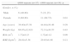
CTG expansion size and cardiovascular risk profileThe correlation between cardiovascular risk and CTG expansion size was assessed by calculating the median number of CTG repeats (900) and dividing the sample into two groups, one with ≤900 and the other with >900 repeats. No significant differences in CTG expansion size were seen in terms of gender, age or BMI (Table 3).
Characterization of the study sample by gender and anthropometric variables according to number of CTG repeats.
| ≤900 repeats (n=15) | >900 repeats (n=16) | p | |
| Gender, n (%) | |||
| Male | 6 (40.00) | 5 (31.25) | |
| Female | 9 (60.00) | 11 (68.75) | 0.61 |
| Age (years) | 39.40±15.38 | 44.44±10.46 | 0.29 |
| Weight (kg) | 69.07±12.021 | 72.31±10.55 | 0.43 |
| BSA (m2) | 1.72±0.13 | 1.72±0.12 | 0.99 |
| BMI (kg/m2) | 26.61±5.36 | 29.45±4.40 | 0.11 |
BMI: body mass index; BSA: body surface area.
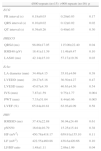
The 12-lead ECG showed no statistically significant differences between the two groups, but patients with >900 repeats had slightly longer PR and QT intervals, and significantly longer QRS duration (0.12±0.02 ms vs. 0.10±0.2 ms; p=0.02) than those with ≤900 repeats.
On high-resolution ECG, patients with larger CTG expansion size had significantly higher mean values for QRSd and LAS40 (Table 4).
Echocardiographic and electrocardiographic characteristics of the study population by number of CTG repeats.
| ≤900 repeats (n=15) | >900 repeats (n=16) | p | |
| ECG | |||
| PR interval (s) | 0.18±0.03 | 0.20±0.03 | 0.17 |
| QRS interval (s) | 0.10±0.02 | 0.12±0.02 | 0.02 |
| QT interval (s) | 0.39±0.26 | 0.40±0.03 | 0.30 |
| HRECG | |||
| QRSd (ms) | 98.86±17.05 | 115.00±22.40 | 0.04 |
| RMS40 (μV) | 18.41±11.39 | 11.49±9.47 | 0.10 |
| LAS40 (ms) | 42.14±15.10 | 57.17±19.36 | 0.03 |
| Echo | |||
| LA diameter (mm) | 34.40±4.15 | 35.81±4.69 | 0.38 |
| LVEDD (mm) | 29.27±5.16 | 30.50±4.27 | 0.47 |
| LVESD (mm) | 45.67±4.30 | 46.63±4.36 | 0.54 |
| IVS (mm) | 7.87±1.59 | 9.75±1.77 | 0.004 |
| PWT (mm) | 7.33±51.04 | 8.44±0.96 | 0.005 |
| LVEF (%) | 65.84±10.81 | 63.93±8.09 | 0.58 |
| HRV | |||
| RMSSD (ms) | 37.43±22.88 | 36.94±24.40 | 0.61 |
| pNN50 | 16.64±16.79 | 15.25±15.44 | 0.38 |
| HF (mV2) | 450.78±418.17 | 659.91±155.10 | 0.11 |
| LF (mV2) | 422.55±460.88 | 438.6±420.66 | 0.10 |
| LF/HF ratio | 1.48±1.11 | 2.06±1.99 | 0.04 |
ECG: electrocardiogram; Echo: echocardiography; HF: high-frequency spectral power; HRECG: high-resolution electrocardiogram; IVS: interventricular septal thickness; LA: left atrial; LAS40: low-amplitude signals (<40 mV); LF: low-frequency spectral power; LVEDD: left ventricular end-diastolic diameter; LVEF: left ventricular ejection fraction; LVESD: left ventricular end-systolic diameter; pNN50: percentage of pairs of NN intervals differing by >50 ms; PWT: posterior wall thickness; QRSd: QRS complex duration; RMS40: root mean square voltage of the last 40 ms of the QRS complex; RMSSD: root mean square of differences of successive NN intervals.
TTE revealed no significant differences between the groups in LA size and IVS thickness, LV end-systolic and end-diastolic diameters, or LVEF, and Holter monitoring also showed no differences.
HRV in patients with >900 repeats (54%; p=0.88) was reduced compared to those with ≤900 repeats (31%; p=0.37).
DiscussionGiven the low prevalence of DM-1, our series is relatively large, representing a significant sample for analysis, which means that our findings may contribute to management of the cardiac aspects of the disease.
Two patients had received permanent pacemakers for symptomatic bifascicular block. None had suffered SCD.
According to the literature, DM-1 is more common in adults, which was the case in our sample, in which most patients were aged between 30 and 40. They were also mostly of short stature and overweight, the latter characteristic possibly being due to the motor limitations and muscle weakness associated with the disease.
With regard to ECG findings, unlike in other series,19,20 the proportion of patients in our study with first-degree AVB was relatively low (38%), but there was a high prevalence of prolonged QRS (51%), in agreement with previous studies,5 indicating significant involvement of the intraventricular conduction system. This may be related to the high proportion (62%) of patients with VLP, which is in line with other studies showing a high prevalence of abnormal findings on high-resolution ECG in these patients (83% of patients with >1000 CTG repeats had VLP in a study by Melacini et al.21), reflecting the existence of areas of slow and fragmented conduction in the myocardium that represent a substrate for reentrant ventricular arrhythmias. However, although this exam is sensitive for the presence of ventricular arrhythmias in DM-1, it has very low specificity, giving a high false positive rate,3 which is corroborated by the fact that no cases of complex or severe ventricular arrhythmias or SCD were recorded in our study population.
We also observed more severe physical repercussions of the disease and worse cardiovascular profile in women, findings that have not been consistently reported in other studies.
With regard to cardiac structural changes in DM-1, there are conflicting reports in the literature, with some authors describing significant echocardiographic alterations, including mitral valve prolapse, LV wall motion abnormalities, reduced LVEF and fractional shortening, LV hypertrophy and dilatation, and systolic dysfunction.12,19,22 No significant echocardiographic alterations were observed in our study population; cardiac dimensions in most patients (94%) were within normal ranges, with slight IVS hypertrophy in 20% and slightly reduced LVEF in one patient.
Another frequent finding in these patients is altered autonomic balance, mainly reflected in vagal dysfunction.5 Our data showed a high proportion of patients with alterations in different HRV parameters, particularly in the frequency domain (LF, HF and LF/HF ratio). Interestingly, female patients presented higher HF and lower LF values and LF/HF ratio than men, reflecting greater vagal activity in women. These results suggest gender-based differences in cardiac autonomic modulation in DM-1, with more severe weakening of vagal activity in men, which may reflect earlier deterioration in baroreceptor function.
Another common subject in the literature is the possible relation between cardiac complications and CTG expansion size in patients with DM-1.5,8,19 This association is supported by the findings of our study, in which first-degree AVB, left bundle branch block, VLP, abnormalities on Holter monitoring, and reduced HRV were significantly associated with larger CTG expansions. We accordingly suggest that CTG expansion size is a valuable tool in cardiovascular risk stratification in these patients, although its real importance remains to be demonstrated in studies with longer follow-up. Although our results did not identify significant arrhythmic risk in this population, patients at risk for disease progression require close monitoring, since the ACC/AHA/ESC guidelines for management of patients with ventricular arrhythmias classify myotonic dystrophy as a class IIb indication for permanent pacemaker implantation.23
The main limitation of the present study is its retrospective nature. Furthermore, although the sample was larger than in most studies on this rare disease, the small number of patients inevitably weakens the statistical power of the analysis.
ConclusionThis case series is the result of a protocol for screening DM-1 patients for heart disease in the cardiology department of Centro Hospitalar Entre Douro e Vouga. The findings of the study show cardiac involvement in DM-1, although the alterations detected were less severe than suggested in the literature. This may reflect the quality of follow-up care offered to these patients in a center with a specialized unit for carriers of this disease.
Ethical disclosuresProtection of human and animal subjectsThe authors declare that the procedures followed were in accordance with the regulations of the relevant clinical research ethics committee and with those of the Code of Ethics of the World Medical Association (Declaration of Helsinki).
Confidentiality of dataThe authors declare that they have followed the protocols of their work center on the publication of patient data.
Right to privacy and informed consentThe authors have obtained the written informed consent of the patients or subjects mentioned in the article. The corresponding author is in possession of this document.
Conflicts of interestThe authors have no conflicts of interest to declare.
Please cite this article as: Gomes L, Pereira T, Martins L. Perfil cardiovascular na distrofia muscular miotónica tipo 1: estudo de uma série de casos seguida num centro especializado. Rev Port Cardiol. 2014;33:765–772.
