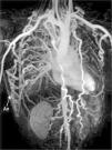
A 22-year-old man was referred to our cardiology department for investigation of hypertension. At presentation his blood pressure was 170/100 mmHg in both arms, and heart rate was 74 beats/min. The pulses were equal over both upper extremities, but bilateral femoral and popliteal pulses were extremely weak and the dorsalis pedis and anterior tibial pulses were not palpable. His past history was unremarkable except for hypertension. There was a II/VI systolic ejection murmur in the left second intercostal area and left scapular region in the back. Transthoracic echocardiography showed concentric left ventricular hypertrophy with normal ventricular function. Magnetic resonance (MR) angiography showed interruption of the descending aorta after the branching of the left subclavian artery (Figure 1). Dilated intercostal, intramammary and thoracodorsal arteries with accompanying anastomosis between the intercostal and thoracodorsal arteries were observed (Figure 2).
Three-dimensional volume rendering magnetic resonance angiography images: (A) left posterior oblique view and (B) right posterior oblique view, showing interruption of the descending aorta after the branching of the left subclavian artery. IA: interrupted aorta; IMA: internal mammary artery; LCCA: left common carotid artery; LSCA: left subclavian artery; RCCA: right common carotid artery; TrBc: truncus brachiocephalicus.
Interrupted aortic arch is defined as a complete luminal and anatomic discontinuity between the ascending and descending aorta. It is a rare and severe congenital heart defect with a very poor prognosis without surgical treatment. Conventional angiography is considered the gold standard and is frequently used in vascular imaging. However, MR imaging with excellent anatomic and functional capabilities can be performed easily, and with advances in imaging technology, acquiring information comparable to that of conventional angiography has become feasible. MR can also be used to assess the vascular anatomy from several views with maximum intensity projection reconstructions.
Ethical disclosuresProtection of human and animal subjectsThe authors declare that no experiments were performed on humans or animals for this study.
Confidentiality of dataThe authors declare that no patient data appear in this article.
Right to privacy and informed consentThe authors declare that no patient data appear in this article.
Conflicts of interestThe authors have no conflicts of interest to declare.


