Journal Information
Vol. 43. Issue 4.
Pages 217-218 (April 2024)
Vol. 43. Issue 4.
Pages 217-218 (April 2024)
Image in Cardiology
Full text access
Acute L waves in heart failure
Ondas Lambda agudas na insuficiência cardíaca
Visits
2921
Juan Diego Sánchez Vega
Corresponding author
jdsanchez@unav.es

Corresponding author.
, Sergio García Gómez, Manuel García de Yébenes Castro
Clínica Universidad de Navarra, Madrid, Spain
This item has received
Article information
Full Text
Bibliography
Download PDF
Statistics
Figures (1)
Additional material (2)
Full Text

L waves are an unusual finding in the analysis of mitral valve pulsed-wave Doppler. This wave is recorded as flow between the left atrium and left ventricle during diastole, due to elevated left ventricular (LV) filling pressures.1–3 In this situation, triphasic mitral filling can be observed, with the presence of mid-diastolic L waves.

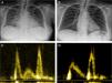
We present the case of a 71-year-old female with preserved LV ejection fraction (LVEF) and moderate ventricular hypertrophy secondary to hypertensive cardiomyopathy, according to a cardiac magnetic resonance study performed the year before. The patient had an episode of acute pulmonary edema during an admission for malaria, with complications of fever, acute kidney disease and hypertensive crisis. During the initial assessment, a transthoracic echocardiogram was performed, which showed high LV filling pressure (E/E′ ratio 23), systolic pulmonary pressure of 60 mmHg estimated by tricuspid regurgitation Doppler, and L waves on mitral pulsed-wave Doppler (Figure 1). Laboratory tests showed NT-proBNP of 1962 pg/ml.

Figure 1.

(A and B) Chest radiography during the episode of heart failure after treatment; (C) L wave between rapid left ventricular (LV) filling (E wave) and atrial contraction (A wave); (D) no L wave after decongestion, with impaired LV relaxation pattern (E

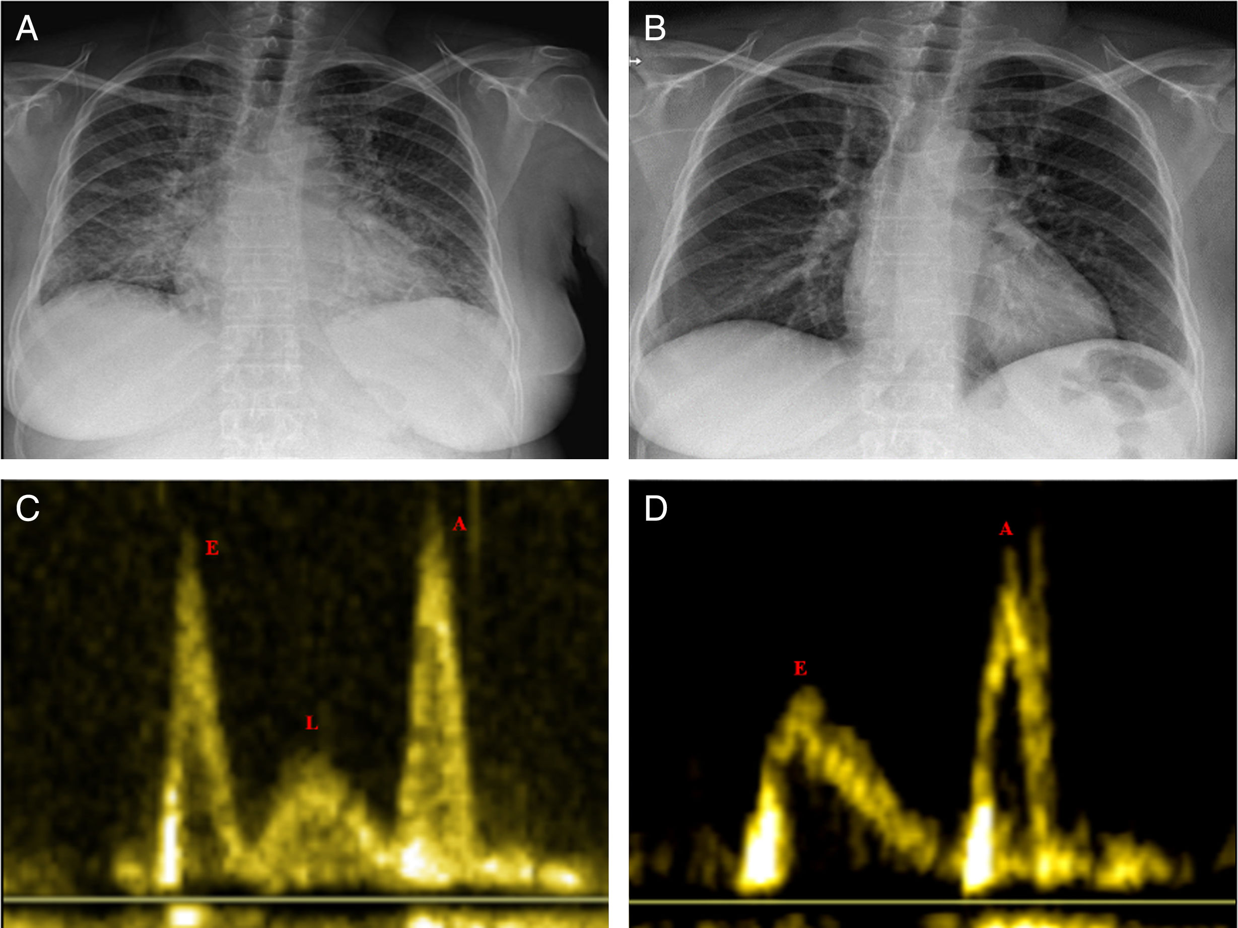
After treatment with antihypertensive drugs and diuretics, following diuresis of 12 l in three days, the patient had normal LV filling pressure (E/E′ ratio 12) and systolic pulmonary pressure of 32 mmHg, and the L wave had disappeared on the echocardiogram, which showed an impaired relaxation pattern in LV filling (EVideos 1 and 2), and a decrease in left atrial volume (from 52.1 to 31.7 ml).

The patient was discharged in good condition after recovery of renal function and adjustment of antihypertensive therapy.

In this paper, we report the presence of L waves during an episode of acute pulmonary edema in a patient with preserved LVEF, in the context of high LV filling pressure. Recognizing this pattern of diastolic filling can be helpful in the rapid diagnosis and treatment of similar cases of heart failure.

Conflicts of interest

The authors have no conflicts of interest to declare.

Appendix A
Supplementary data

The following are the supplementary data to this article:

(0.11MB)
(0.09MB)

References
[1]
V. Kumar, J. Jose, V.J. Jose.
L wave in echo Doppler.
Indian Heart J, 66 (2014), pp. 392-393
[2]
C.S.P. Lam, L. Han, J.W. Ha, et al.
The mitral L wave: a marker of pseudonormal filling and predictor of heart failure in patients with left ventricular hypertrophy.
J Am Soc Echocardiogr, 18 (2005), pp. 336-341
[3]
J.W. Ha, J.A. Ahn, J.Y. Moon, et al.
Triphasic mitral inflow velocity with mid-diastolic flow: the presence of mid-diastolic mitral annular velocity indicates advanced diastolic dysfunction.
Eur J Echocardiogr, 7 (2006), pp. 16-21
Copyright © 2023. Sociedade Portuguesa de Cardiologia
Download PDF
Idiomas
Revista Portuguesa de Cardiologia (English edition)
Article options
Tools
Supplemental materials
en pt

Are you a health professional able to prescribe or dispense drugs?

Você é um profissional de saúde habilitado a prescrever ou dispensar medicamentos

By checking that you are a health professional, you are stating that you are aware and accept that the Portuguese Journal of Cardiology (RPC) is the Data Controller that processes the personal information of users of its website, with its registered office at Campo Grande, n.º 28, 13.º, 1700-093 Lisbon, telephone 217 970 685 and 217 817 630, fax 217 931 095, and email revista@spc.pt. I declare for all purposes that the information provided herein is accurate and correct.