Informação da revista
Vol. 35. Núm. 3.
Páginas 187-188 (Março 2016)
Partilhar
Partilhar
Baixar PDF
Mais opções do artigo
Vol. 35. Núm. 3.
Páginas 187-188 (Março 2016)
Image in Cardiology
Open Access
Acquired mitral valve aneurysm due to severe aortic regurgitation
Aneurisma na válvula mitral devido a regurgitação aórtica grave
Visitas
6694
Rezvaniyeh Salehia, Leili Pourafkaria,b,
Autor para correspondência
leili.p@gmail.com

Corresponding author.
, Nader Djalal Naderb
a Cardiovascular Research Center, Tabriz University of Medical Sciences, Tabriz, Iran
b University at Buffalo, Buffalo, NY, United States
Este item recebeu

Under a Creative Commons license
Informação do artigo
Texto Completo
Baixar PDF
Estatísticas
Figuras (4)
Mostrar maisMostrar menos
Material adicional (2)
Texto Completo

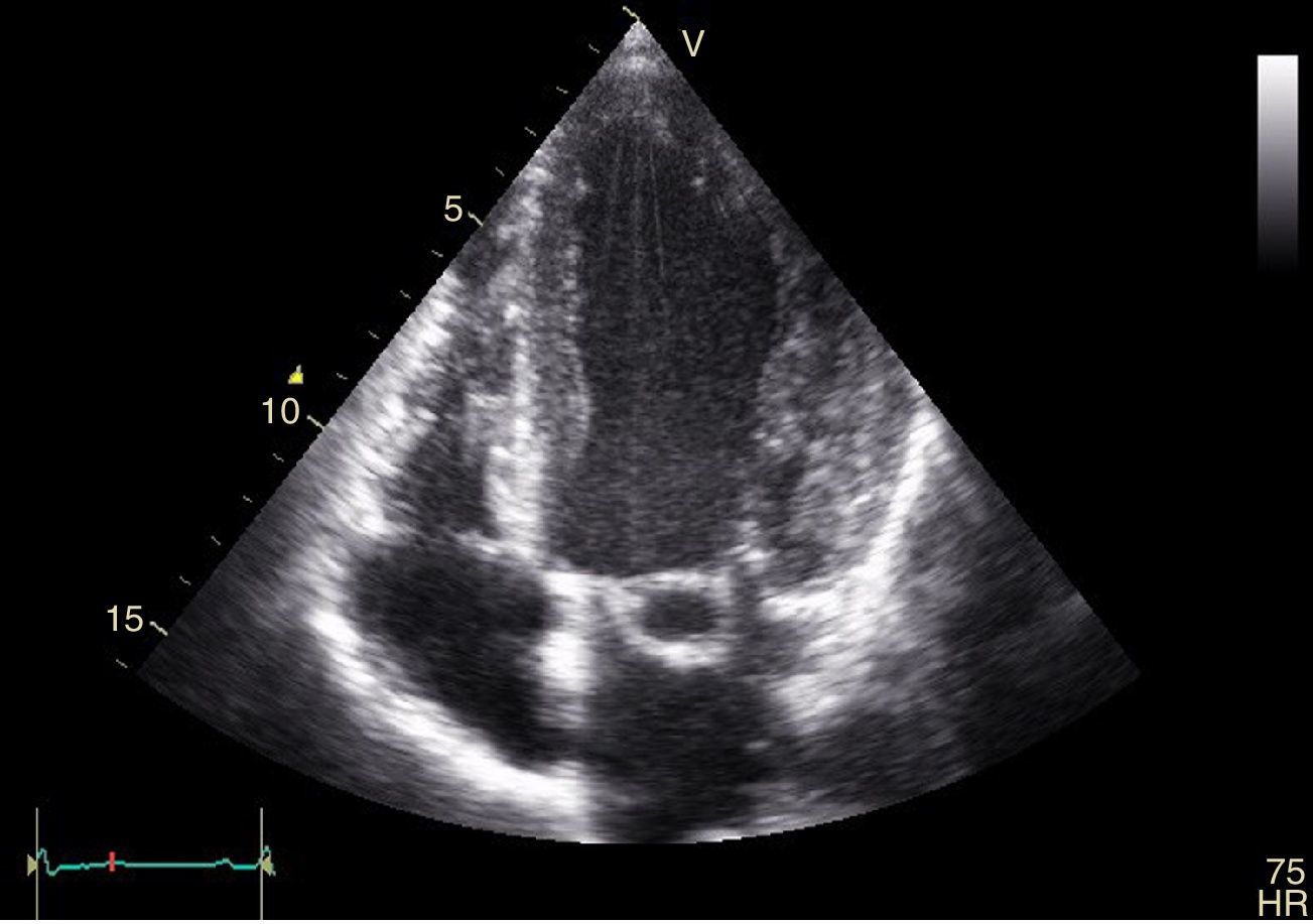
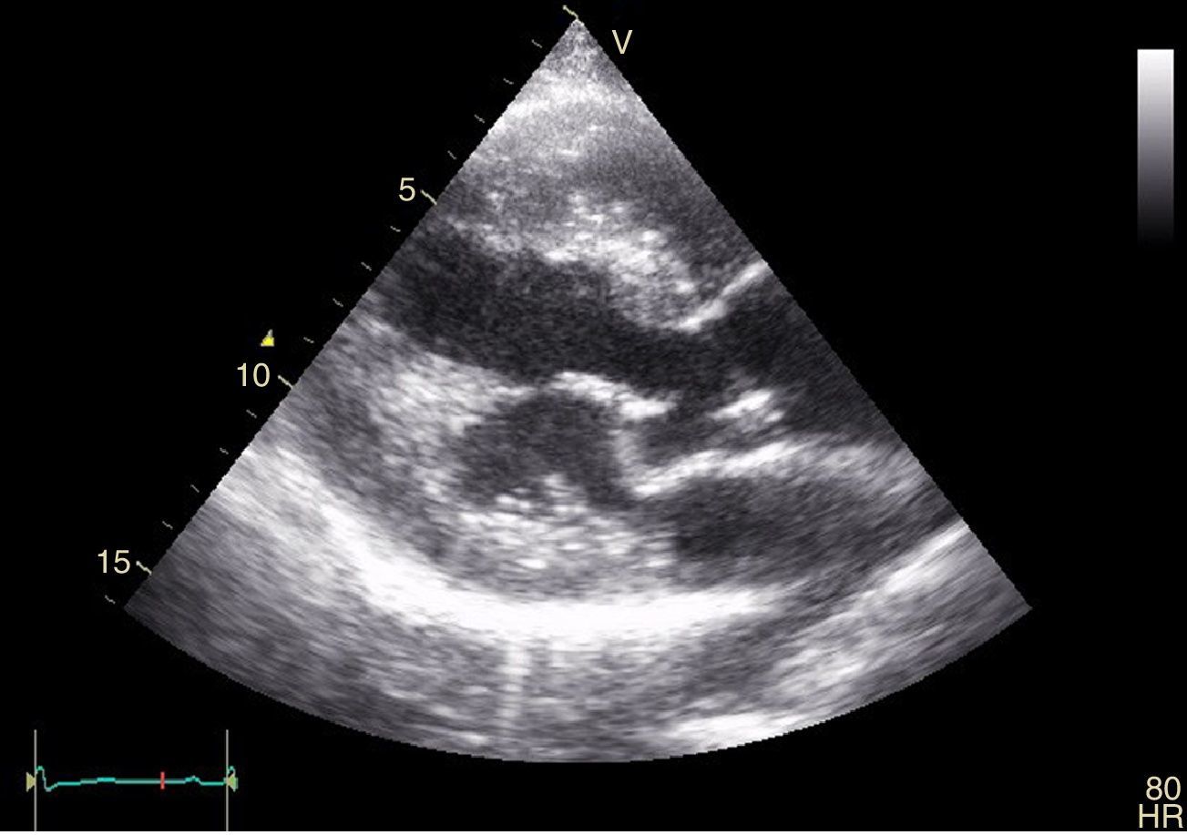
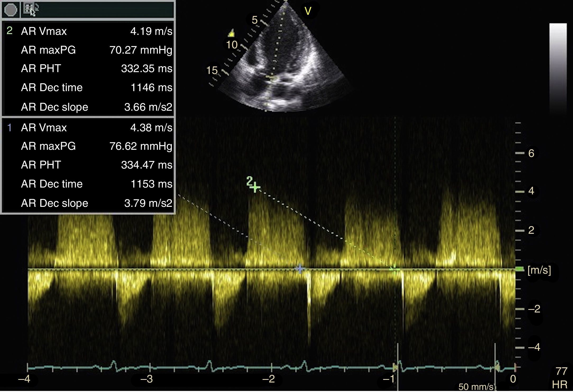
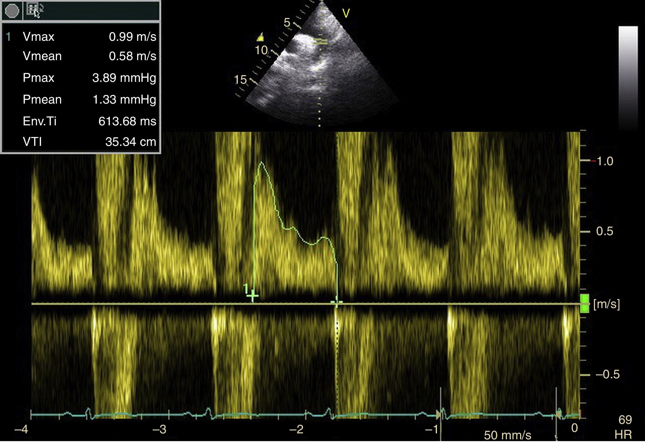
A 52-year-old man presented to our emergency department with shortness of breath. He reported exertional dyspnea for the past five years which had worsened recently. He reported no weight loss, fever or poor appetite. His past medical history was noncontributory. The physical exam revealed a diastolic murmur best heard in the aortic area. Echocardiography showed a mildly enlarged left ventricle with normal contraction. Severe aortic regurgitation (AR) and holodiastolic flow reversal in the abdominal aorta were present (Figures 1–4). No vegetation was seen on the aortic valve. There was an aneurysmal sac on the anterior leaflet of the mitral valve, corresponding to the site of the high velocity AR jet (video 1 and video 2 in the Supplementary material). Laboratory tests including complete blood cell count, liver and kidney function tests, erythrocyte sedimentation rate and C-reactive protein were normal. The patient refused surgical intervention and was lost to follow-up.

Figure 1.

Transthoracic echocardiogram, 4-chamber apical view, showing the aneurysmal sac of the anterior mitral valve leaflet.

(0,09MB).
Figure 2.

Transthoracic echocardiogram, long-axis parasternal view, showing the mitral valve anterior leaflet aneurysm.

(0,09MB).
Figure 3.

Continuous wave spectral Doppler recording through the aortic valve. Pressure half time is noted to be 334 ms.

(0,26MB).
Figure 4.

Holodiastolic flow reversal in the descending thoracic aorta indicative of significant aortic valve regurgitation.

(0,29MB).

Acquired mitral valve aneurysm is a rare entity which has generally been reported in the context of endocarditis. Aneurysm of the anterior mitral valve leaflet due to the regurgitant flow of AR is a plausible mechanism in the absence of evidence for endocarditis.

Ethical disclosuresProtection of human and animal subjects

The authors declare that no experiments were performed on humans or animals for this study.

Confidentiality of data

The authors declare that they have followed the protocols of their work center on the publication of patient data.

Right to privacy and informed consent

The authors have obtained the written informed consent of the patients or subjects mentioned in the article. The corresponding author is in possession of this document.

Conflicts of interest

The authors have no conflicts of interest to declare.

Appendix A
Supplementary material

The following are supplementary material to this article:

(0,25MB)
(0,27MB)

Copyright © 2016. Sociedade Portuguesa de Cardiologia
Idiomas
Revista Portuguesa de Cardiologia
Opções de artigo
Ferramentas
Material Suplementar
en pt

Are you a health professional able to prescribe or dispense drugs?

Você é um profissional de saúde habilitado a prescrever ou dispensar medicamentos

Ao assinalar que é «Profissional de Saúde», declara conhecer e aceitar que a responsável pelo tratamento dos dados pessoais dos utilizadores da página de internet da Revista Portuguesa de Cardiologia (RPC), é esta entidade, com sede no Campo Grande, n.º 28, 13.º, 1700-093 Lisboa, com os telefones 217 970 685 e 217 817 630, fax 217 931 095 e com o endereço de correio eletrónico revista@spc.pt. Declaro para todos os fins, que assumo inteira responsabilidade pela veracidade e exatidão da afirmação aqui fornecida.