
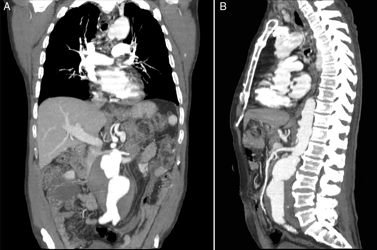
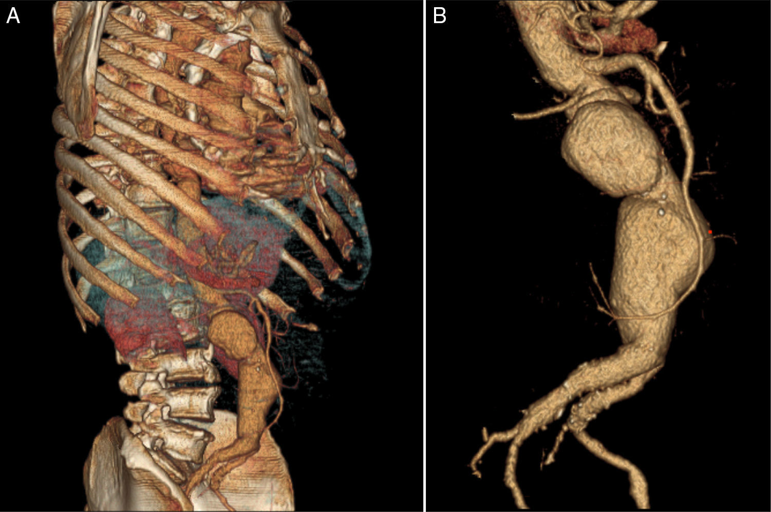
Abdominal aortic aneurysm is a relatively common condition associated with well-defined cardiovascular risk factors. A 74-year-old Caucasian man with a 100-pack-year history of cigarette smoking and generalized atherosclerotic disease manifested by symptomatic carotid artery disease and peripheral artery disease presented to our department for assessment of severe abdominal pain. Physical examination was unremarkable, except for a tender and pulsatile periumbilical mass. A contrast-enhanced computed tomography scan showed an aortic aneurysm with circumferential mural thrombosis extending from below the renal arteries to the bifurcation of the common iliac arteries, without evidence of rupture or dissection (Figure 1A and B). Three-dimensional angiographic reconstruction revealed a complex bilobular aneurysm, the proximal and distal segments measuring 6.6 cm×5.6 cm and 7.5 cm×6.8 cm, respectively, in transverse diameters (Figure 2A and B; Supplementary Data, Videos 1 and 2). The patient underwent elective surgery three months later, with successful aorto-aortic graft insertion.
 and sagittal view (B).")
 and corresponding volume-rendered angiography in detail (B).")
This case illustrates the usefulness of three-dimensional computed tomography angiography in the assessment of the location and anatomy of aortic aneurysms, providing a valuable non-invasive tool for selecting the appropriate surgical procedure. The rare morphology of this aneurysm, in particular, resembles that of a human fetus in the uterus, giving it an unusual and curious radiological appearance.
Ethical disclosuresProtection of human and animal subjectsThe authors declare that no experiments were performed on humans or animals for this study.
Confidentiality of dataThe authors declare that they have followed the protocols of their work center on the publication of patient data.
Right to privacy and informed consentThe authors declare that no patient data appear in this article.
Conflicts of interestThe authors have no conflicts of interest to declare.
