
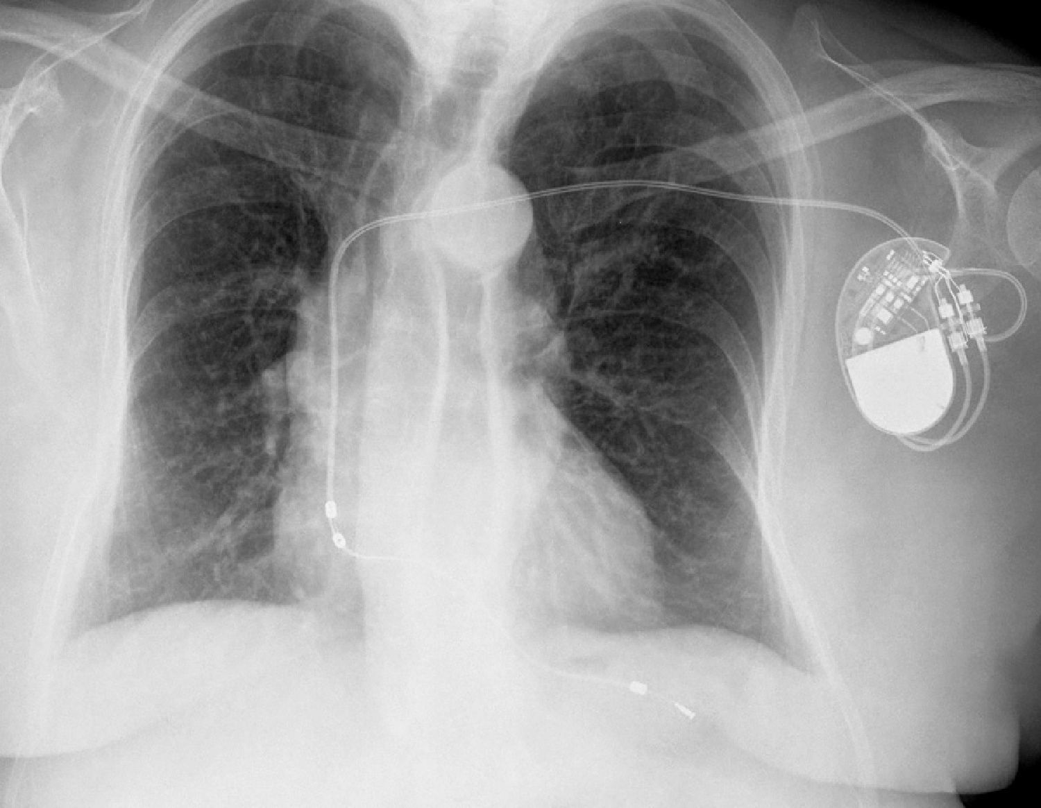
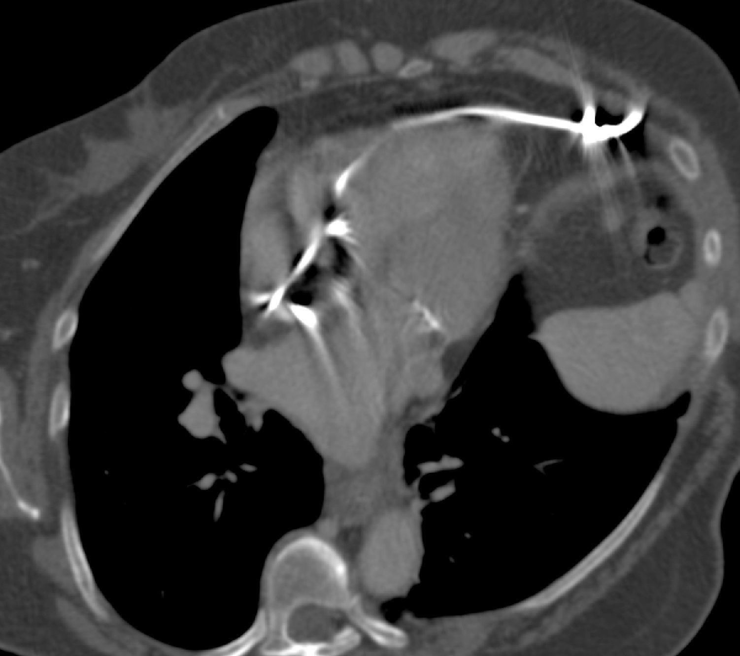
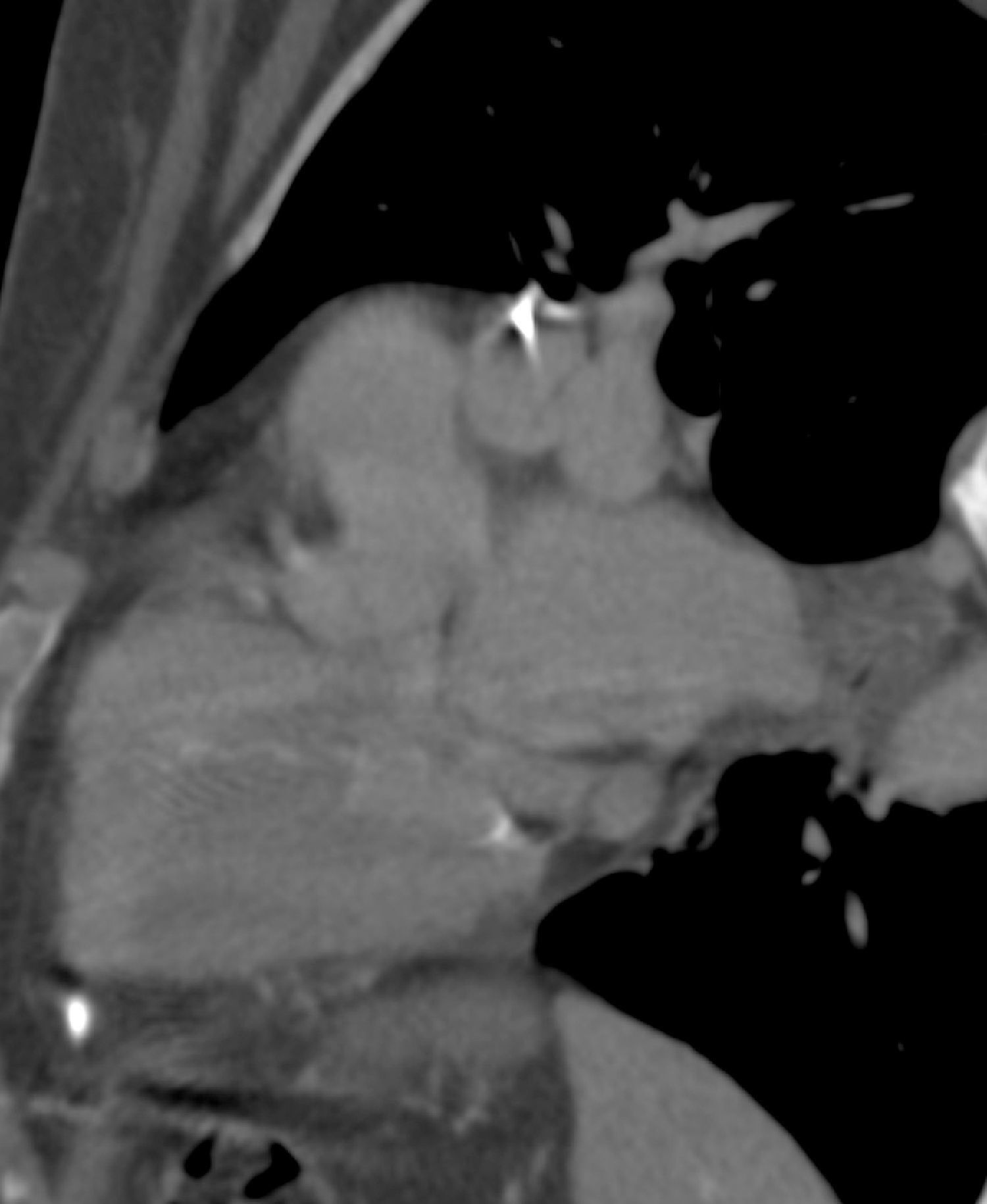

A 74-year-old woman had undergone implantation of a permanent pacemaker (VDD) for grade 3 atrioventricular block two years previously, with no intervening complications. At a routine follow-up consultation, although she presented no symptoms, pacemaker dysfunction was detected (undersensing and failure to capture). A posteroanterior chest X-ray showed the pacemaker lead tip protruding outside the heart (Figure 1). Thoracic 16-slice multidetector computed tomography, with multiplanar and three-dimensional reconstructions, confirmed the position of the lead tip outside the heart, with an intrathoracic course towards the diaphragm (Figures 2–4).


Multidetector computed tomography, with multiplanar reconstruction in oblique axial view, showing the lead course, initially intraventricular and then passing through the right ventricular free wall, with its tip in the paracardiac fat on the left side, close to the chest wall. No pericardial effusion or significant changes in mediastinal fat were observed.


Perforation of the myocardium by a pacemaker lead is a major, albeit uncommon, complication of such devices, with an incidence of 0.3–1.2%.
Perforations are classified as acute (5–7 days after device implantation), subacute (7–30 days) or late (more than 30 days). Most cases reported in the literature occurred in the first year.
Most patients with myocardial perforation are symptomatic, with chest pain, dyspnea, hypotension or pacemaker dysfunction. Occasionally there may be symptoms triggered by stimulation of the chest wall muscles or hiccups through stimulation of the diaphragm. More rarely, as in the present case, the patient can be completely asymptomatic, and a high level of suspicion is required. Conventional X-ray and computed tomography can confirm the diagnosis and determine the lead course.
Ethical disclosuresProtection of human and animal subjectsThe authors declare that no experiments were performed on humans or animals for this study.
Confidentiality of dataThe authors declare that no patient data appear in this article.
Right to privacy and informed consentThe authors declare that no patient data appear in this article.
Conflicts of interestThe authors have no conflicts of interest to declare.
Please cite this article as: Valentim MH, et al. Perfuração tardia do miocárdio por cateter de pacemaker em doente assintomática. Rev Port Cardiol. 2013. http://dx.doi.org/10.1016/j.repc.2012.10.009
