
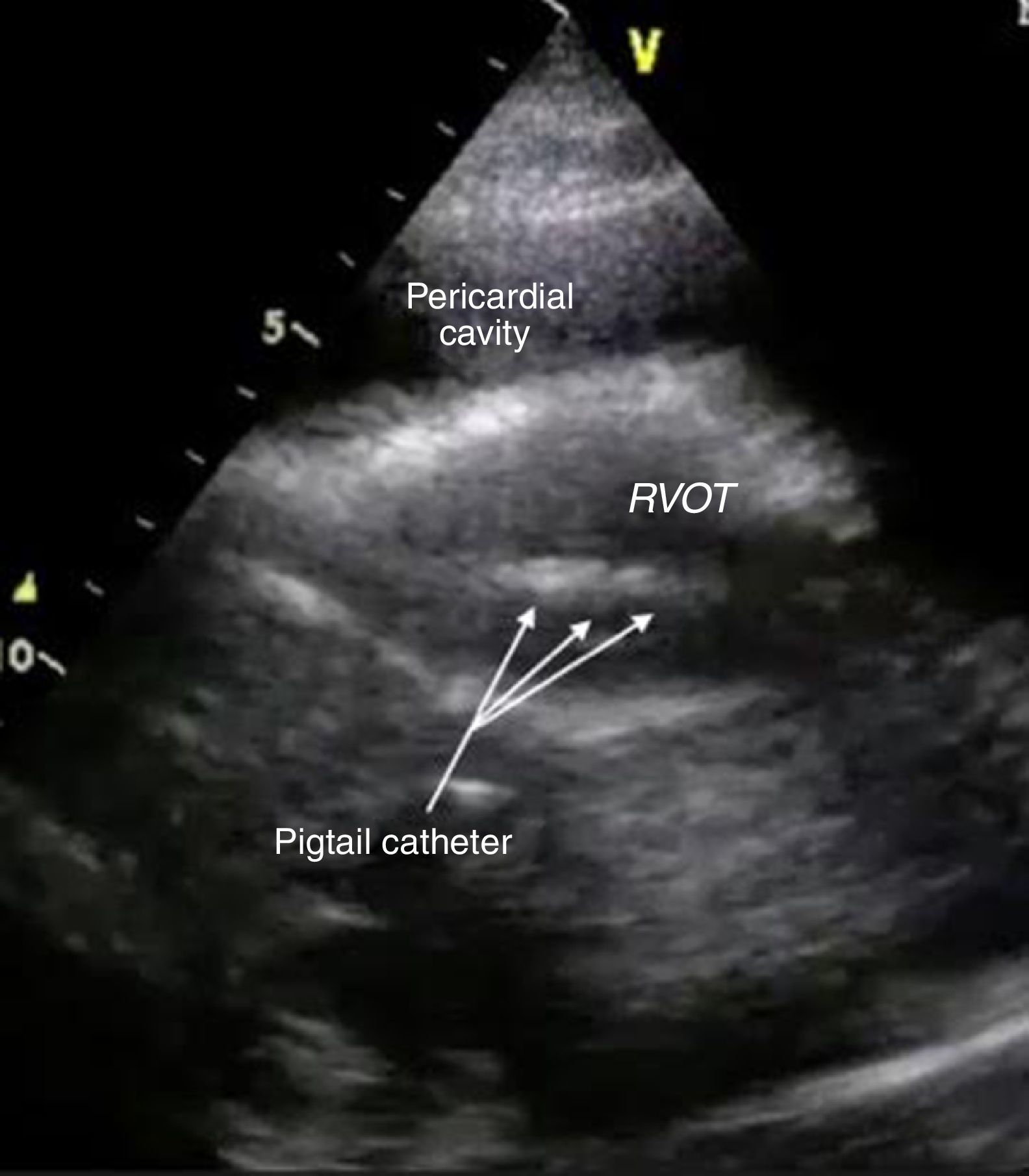
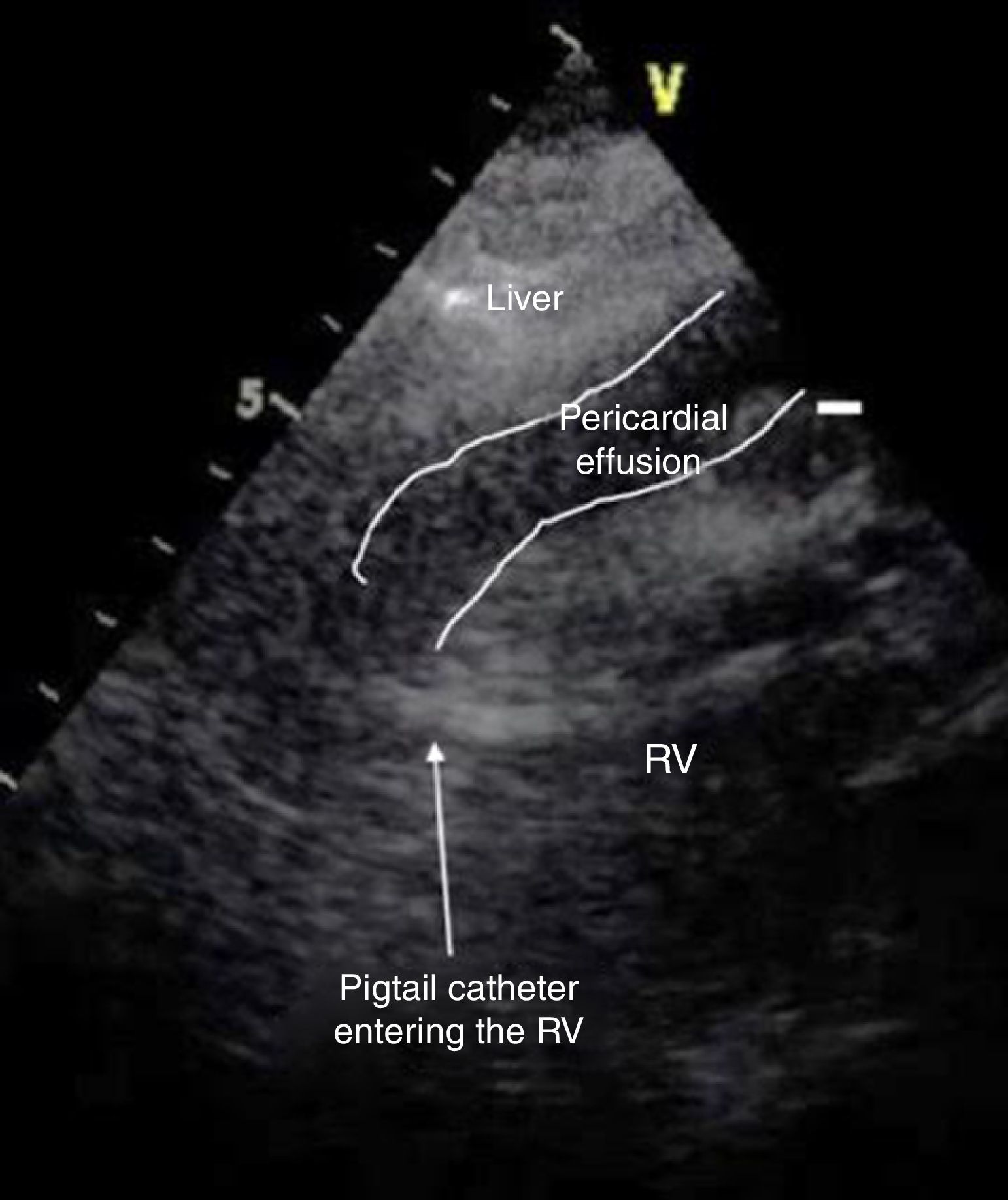
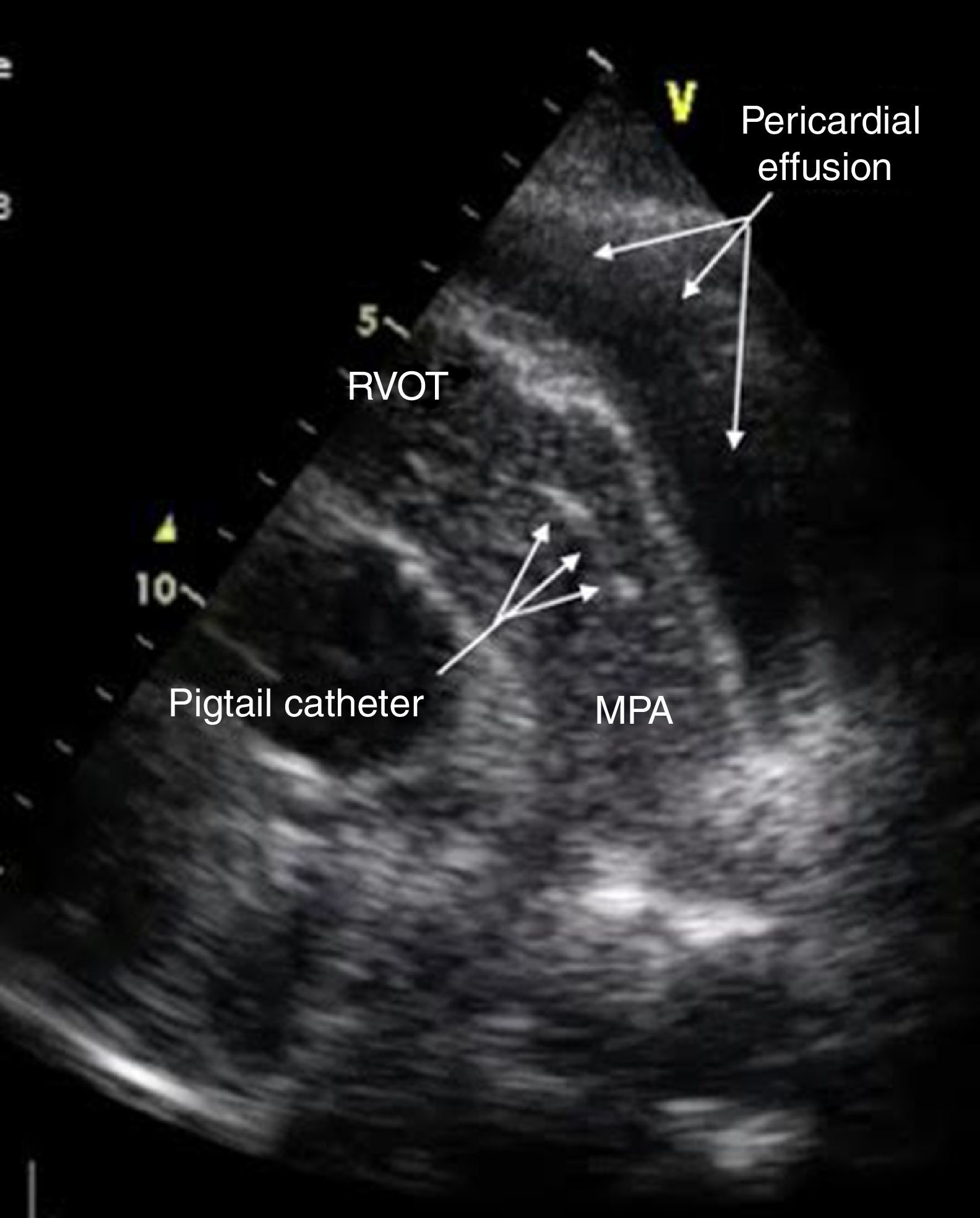
A 77-year-old woman presented to our emergency department with worsening shortness of breath. She was hemodynamically stable. Initial work-up was significant for hemoglobin 10.3 g/dl and enlarged cardiac silhouette on the chest X-ray. Transthoracic echocardiography showed a large circumferential pericardial effusion and dilated inferior vena cava. There was no right chamber collapse. Therapeutic pericardiocentesis was performed in the cardiac catheterization laboratory. An attempt was made to drain the pericardial fluid through a subxiphoid window under fluoroscopic guidance using a percutaneous needle. About 200 ml of serosanguinous fluid was aspirated. A guide wire was then advanced through the needle hub, the needle was removed and a pigtail catheter was introduced and left in place through a catheter-over-wire technique for further drainage. The procedure was considered successful by the performing cardiologist and the patient was admitted. No further drainage was noticed during the next 12 hours. The patient remained stable but did not report any improvement in her symptoms. Follow-up echocardiography 12 hours after the procedure showed massive pericardial effusion with dilated inferior vena cava. The catheter was seen in the right ventricle passing through the right ventricular free wall (Figures 1 and 2). Intravenous injection of agitated saline showed the catheter had advanced to the main pulmonary artery (Figure 3, Videos 1-3). Inadvertent right ventricular perforation and advancement of the pigtail catheter to the pulmonary artery was diagnosed and the patient required surgery for removal of the catheter and repair of the right ventricular free wall.


The case highlights a potentially serious complication of fluoroscopy-guided pericardiocentesis. Echocardiography-guided pericardiocentesis is shown to be safe and effective, and is the technique of choice for draining pericardial effusions. Additionally, initial drainage should be done though the catheter using an over-the-wire technique rather than needle aspiration, which should be reserved for emergent situations and limited access to imaging modalities.
Ethical disclosuresProtection of human and animal subjectsThe authors declare that no experiments were performed on humans or animals for this study.
Confidentiality of dataThe authors declare that no patient data appear in this article.
Right to privacy and informed consentThe authors declare that no patient data appear in this article.
Conflicts of interestThe authors have no conflicts of interest to declare.
