
Constrictive pericarditis is a rare disease, usually the result of long-standing pericardial inflammation leading to pericardial scarring with thickening, fibrosis, and calcification. The most frequent causes are mediastinal radiation, chronic idiopathic pericarditis, cardiac surgery, and tuberculous pericarditis. Nearly 9% of patients with acute pericarditis for any reason go on to develop constrictive physiology.
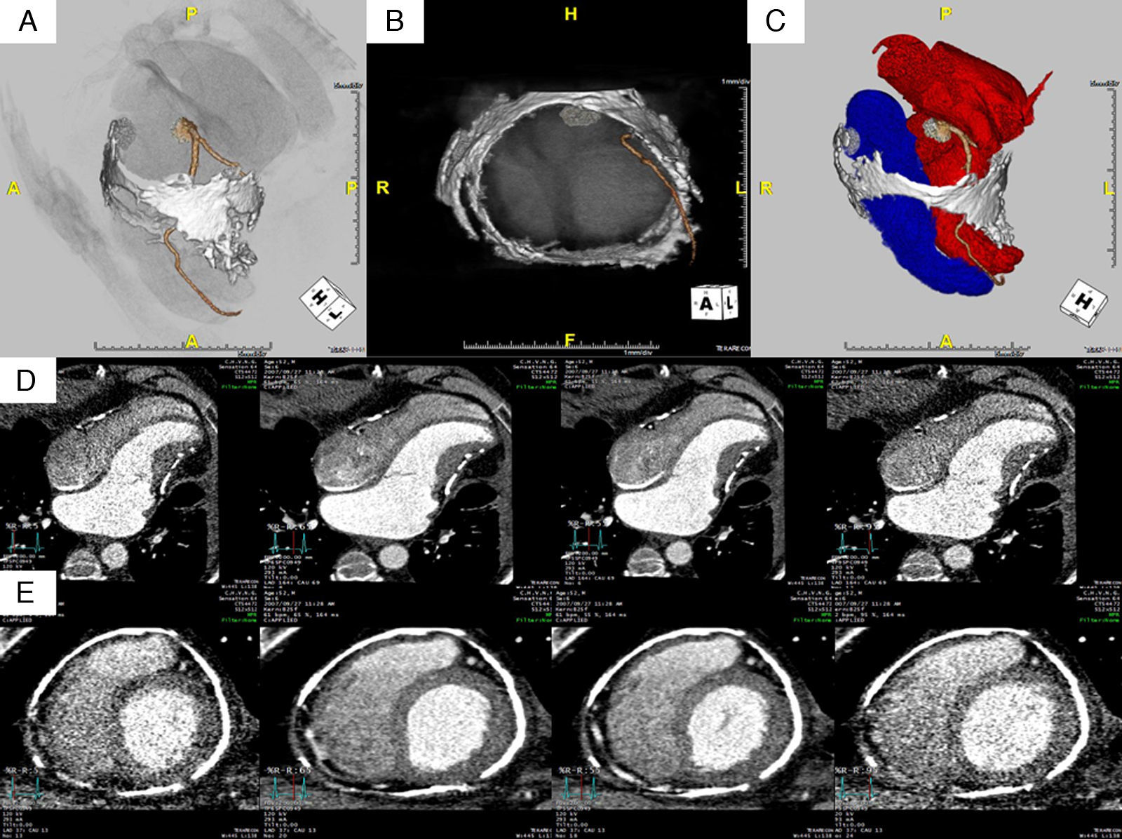
We report the case of a 52-year-old man with a history of acute pericarditis 30 years ago, with a three-year evolution of fatigue, dyspnea and palpitations. Physical examination revealed jugular venous distension and hepatomegaly and a pericardial knock was audible on cardiac auscultation. The 12-lead electrocardiogram revealed atrial flutter and the transthoracic echocardiogram showed distorted left ventricular geometry. Cardiac computed tomography (CT) demonstrated pericardial thickening and extensive pericardial calcification (Figure 1A,B and C), mostly mid-cardiac, causing distortion of left ventricular morphology. Marked restriction to diastolic ventricular filling was observed, with diastolic apical dilatation (Figure 1D and E). Cardiac catheterization showed ventricular pressure waveforms with the classic dip-and-plateau physiology and equalization of end-diastolic pressures of both ventricles, compatible with pericardial constriction.
 showing the thickened and calcified pericardium and its relations with the coronary arteries and cardiac chambers. Maximum intensity projection images in long- (D) and short-axis (E) views in different phases of the cardiac cycle illustrate restricted myocardial movement and diastolic apical dilatation. Tube current modulation was used to reduce radiation exposure, with full current applied only in diastolic phases (note differences in image quality in systole and diastole).")
Multidetector computed tomography post-processed volume rendered images (A, B and C) showing the thickened and calcified pericardium and its relations with the coronary arteries and cardiac chambers. Maximum intensity projection images in long- (D) and short-axis (E) views in different phases of the cardiac cycle illustrate restricted myocardial movement and diastolic apical dilatation. Tube current modulation was used to reduce radiation exposure, with full current applied only in diastolic phases (note differences in image quality in systole and diastole).
This case highlights the role of cardiac CT as an appropriate imaging technique for the diagnosis of constrictive pericarditis.
Ethical disclosuresProtection of human and animal subjectsThe authors declare that no experiments were performed on humans or animals for this investigation.
Confidentiality of dataThe authors declare that no patient data appear in this article.
Right to privacy and informed consentThe authors declare that no patient data appear in this article.
Conflicts of interestThe authors have no conflicts of interest to declare.
