
Acute type A aortic dissection is a surgical emergency, and the development of false aneurysms is one reason for reoperation.
An 82-year-old man went to the emergency department with oppressive chest pain radiating to the back for three weeks and symptoms of heart failure, in NYHA class III/IV. He had a history of acute type A aortic dissection and had had a Dacron tube graft implanted at the age of 64.
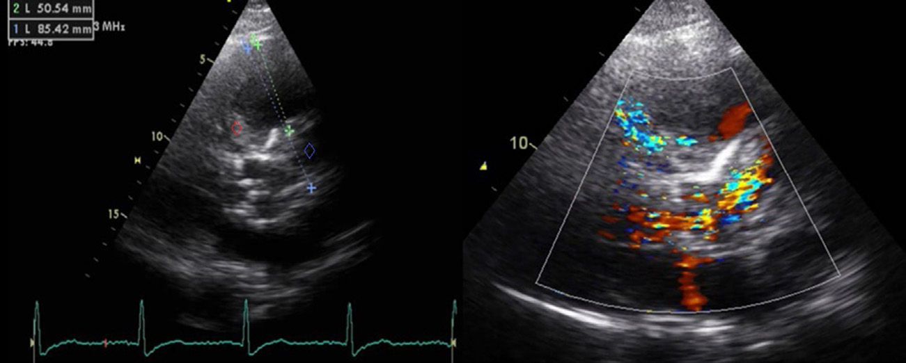
The transthoracic echocardiogram (Figure 1) showed the Dacron graft in the ascending aorta and an apparent solution of continuity in the proximal portion of the anastomosis, a large aneurysm (53 mm × 71 mm) with thrombus compressing the pulmonary artery trunk (peak gradient 27 mmHg), and moderate to severe aortic regurgitation.
 and a large aneurysm (53 mm × 71 mm) with thrombus (red diamond) and flow apparently originating from the proximal and distal ends of the tube.")
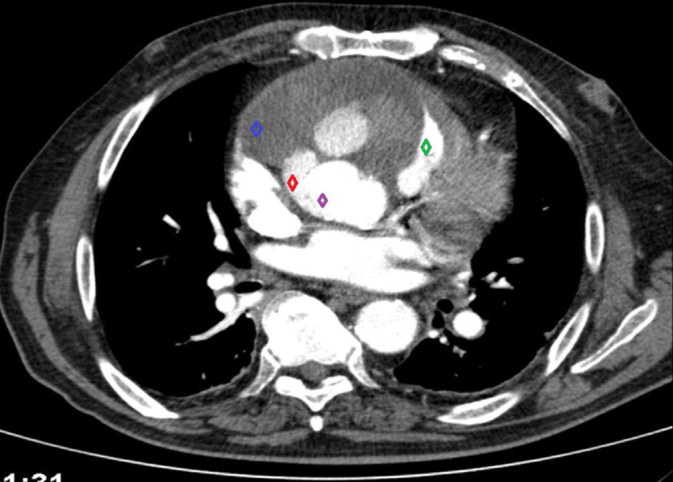
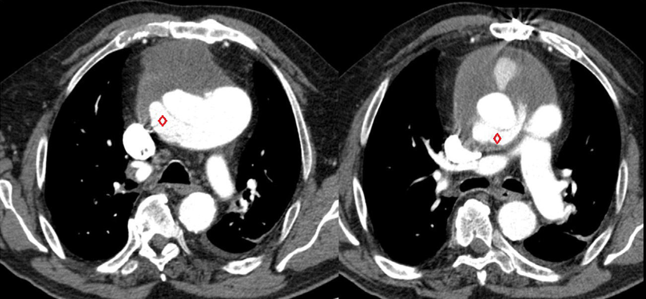
Thoracic computed tomography angiography (Figures 2 and 3) showed the Dacron tube from 2.1 cm above the aortic valve leaflets to 1.8 cm after the emergence of the brachiocephalic trunk surrounded by an image suggestive of an ascending aortic aneurysm (10 cm × 10 cm) with two active contrast leaks at the ends of the graft (type 1 endoleak), compressing the right ventricle, pulmonary artery trunk and right coronary artery.
 due to a proximal leak from the Dacron tube (purple diamond) and a large ascending aorta aneurysm with organized thrombus (blue diamond) compressing the pulmonary artery trunk (green diamond).")
 due to a distal leak from the Dacron tube (red diamond) (type 1 endoleak).")
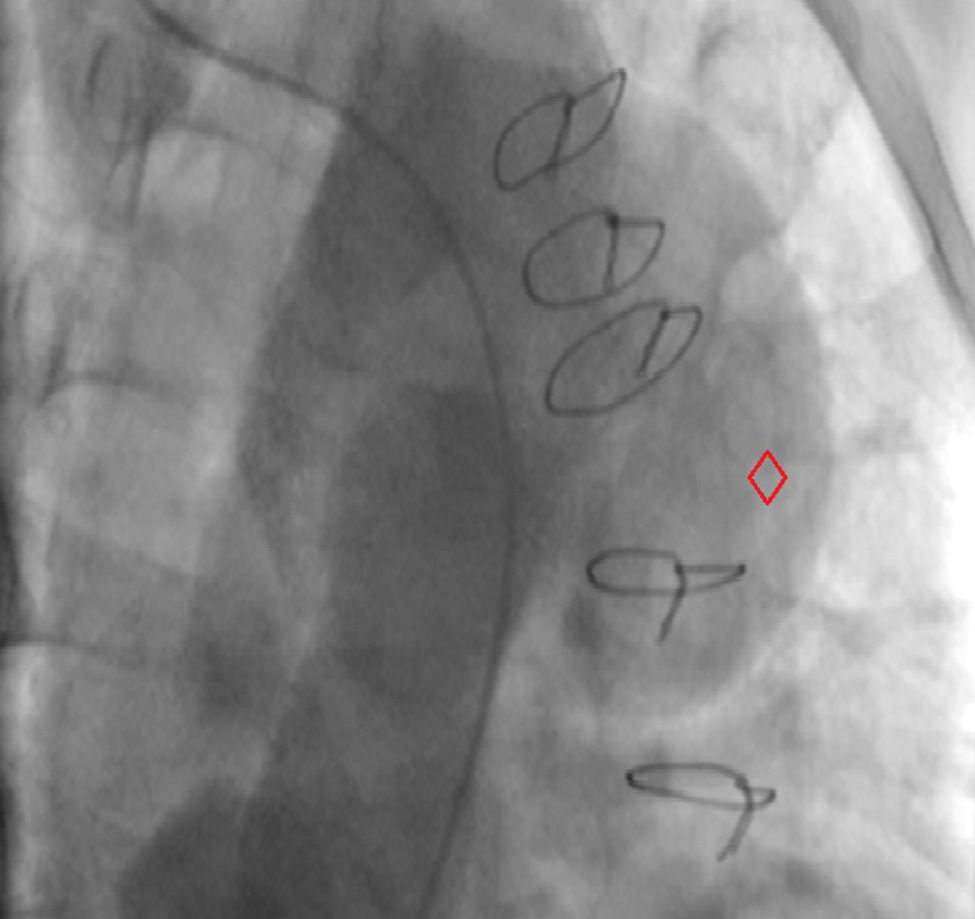
Aortography (Figure 4) revealed an apparent proximal leak in the Dacron tube and a false lumen forming a large saccular aneurysm.
.")
Following joint medical and surgical evaluation, it was decided that the patient was unsuitable for cardiac surgery due to his multiple comorbidities (chronic obstructive pulmonary disease, obstructive sleep apnea syndrome and chronic renal failure), which was also in agreement with his and his family's wishes. He was discharged in NYHA class II–III/IV.
The patient was readmitted four months later due to heart failure and died from a nosocomial infection.
Conflicts of interestThe authors have no conflicts of interest to declare.
Please cite this article as: Caetano F, Fernandes A, Trigo J, Basso S, Botelho A, Leitão Marques A. Dissecção aórtica tipo A aguda corrigida: caso encerrado? Rev Port Cardiol. 2013;32:737–739.
