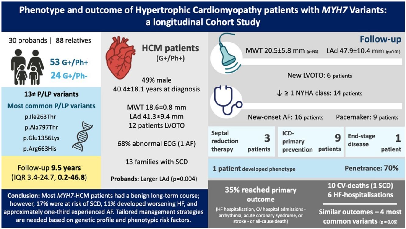

Hypertrophic cardiomyopathy (HCM) is primarily caused by mutations in the MYH7 and MYBPC3 genes which exhibit diverse clinical expression and prognosis. This study aims to outline the clinical features and long-term cardiovascular outcome of patients with MYH7-related HCM, and to look for clinical and prognostic implications of specific variants.
MethodsA retrospective longitudinal analysis was conducted on 30 unrelated HCM families with pathogenic/likely pathogenic (P/LP) MYH7 mutations. A composite endpoint encompassing hospitalisation for heart failure, cardiovascular admissions, or all-cause mortality was evaluated. Kaplan–Meier survival analysis was used to compare outcomes across prevalent variants and genetic profiles.
ResultsAmong 118 individuals (30 probands, 88 relatives), 77 were carriers of P/LP MYH7 variants – 69% with HCM (G+/Ph+) at diagnosis and 31% P/LP MYH7 variant carriers only (G+/Ph−). Thirteen different P/LP variants were identified in the 30 families, with four (p.Ile263Thr, p.Ala797Thr, p.Glu1356Lys, p.Arg663His) accounting for two-thirds of cases. HCM patients had a mean age at diagnosis of 40.4±18.1 years and 49% were male. Baseline maximal wall thickness (MWT) was 18.6±0.8 mm, left atrial diameter (LAD) was 41.3±9.4 mm, and 23% exhibited resting left ventricular outflow tract obstruction (LVOTO); 68% had abnormal ECGs, but only one patient had atrial fibrillation (AF). Probands showed significantly larger LAD than relatives (p=0.004). A history of premature familial sudden cardiac death (SCD) was reported in 43% families. During a median follow-up of 9.5 years (IQR 3.4–24.7, range 0.2–46.8 years), 35% of patients reached the composite endpoint, including 16 deaths (1 SCD) and 6 heart failure (HF)-related hospitalisations; 30% of patients developed AF, 17% received an ICD for primary prevention of SCD and 17% had a pacemaker implantation. Older age at diagnosis (p<0.01) and increased LAD (p=0.048) predicted poorer outcomes. The composite endpoint was similar between probands and relatives with HCM (p=0.08) and across the main variants (p=0.06). Overall penetrance was 70%, with only one carrier progressing to mild HCM. ESC HCM risk-SCD scores were similar across variants, both at baseline (p=0.601) and at last follow-up (p=0.286).
ConclusionsMost patients with MYH7-related HCM presented with a benign phenotype over the long term. Nonetheless, the risks of AF, SCD, and worsening HF throughout life justify regular monitoring, and the need to look for particular genetic profiles that may potentially help tailored management strategies.
A miocardiopatia hipertrófica (MCH) é causada maioritariamente por variantes patogénicas nos genes MYH7 e MYBPC3, apresentando expressão clínica e prognóstico heterogéneos. Este estudo teve como objetivo caracterizar as manifestações clínicas e os desfechos cardiovasculares a longo prazo de doentes com MCH associada a variantes no gene MYH7, bem como avaliar as implicações clínicas e prognósticas de variantes específicas.
MétodosRealizou-se um estudo retrospetivo longitudinal que incluiu 30 famílias não relacionadas afetadas com MCH e portadoras de variantes patogénicas ou provavelmente patogénicas (P/LP) no gene MYH7. O desfecho composto incluiu hospitalização por insuficiência cardíaca, otras hospitalizações de causa cardiovascular ou morte por todas as causas. A sobrevivência foi analisada pelo método de Kaplan–Meier para comparar os desfechos entre as variantes mais prevalentes.
ResultadosDos 118 indivíduos estudados (30 probandos, 88 familiares), 77 eram portadores de variantes P/LP no gene MYH7 — 69% apresentavam MCH (G+/Ph+) ao diagnóstico e 31% eram apenas portadores (G+/Ph−). Foram identificadas 13 variantes P/LP distintas, sendo que quatro delas (p.Ile263Thr, p.Ala797Thr, p.Glu1356Lys, p.Arg663His) representavam dois terços dos casos. Os doentes com MCH tinham uma idade média ao diagnóstico de 40,4±18,1 anos e 49% eram do sexo masculino. A espessura parietal máxima (MWT) aquando da primeira avaliação era de 18,6±0,8mm, o diâmetro da aurícula esquerda (AEd) era de 41,3±9,4mm, e 23% apresentavam obstrução do trato de saída do ventrículo esquerdo (TSVE) em repouso; 68% apresentavam alterações no ECG, e apenas um doente tinha fibrilhação auricular (FA). Os probandos apresentavam um AEd significativamente maior do que os seus familiares (p=0,004). Uma história familiar de morte súbita cardíaca (MSC) foi documentada em 43% das famílias. Durante um seguimento mediano de 9,5 anos (IIQ 3,4–24,7; 0,2–46,8 anos), 35% dos doentes atingiram o desfecho composto, incluindo 16 óbitos (1 MSC) e 6 hospitalizações por insuficiência cardíaca. Durante o seguimento, 30% desenvolveram FA, 17% receberam um cardioversor-desfibrilhador implantável (CDI) para prevenção primária e 17% foram submetidos à implantação de pacemaker. A idade mais avançada ao diagnóstico (p<0,01) e o AEd aumentado (p=0,048) associaram-se a desfechos desfavoráveis. O desfecho composto foi semelhante entre probandos e familiares com MCH (p=0,08) e entre as variantes mais prevalentes (p=0,06). A penetrância global foi de 70%, com apenas um portador a desenvolver o fenótipo de MCH durante o seguimento. O escore ESC HCM Risk-SCD foi semelhante entre as variantes, tanto na avaliação inicial (p=0,601) como no final do seguimento (p=0,286).
ConclusõesA maioria dos doentes com MCH associada a variantes P/LP no gene MYH7 apresentou um fenótipo benigno durante um longo período de seguimento. No entanto, a ocorrência de FA, MSC e progressão para insuficiência cardíaca ao longo da vida justifica uma monitorização regular, assim como a necessidade de investigar perfis genéticos específicos que possam, futuramente, apoiar estratégias de gestão personalizada.

Hypertrophic cardiomyopathy (HCM) is a heart muscle disease characterized by unexplained left ventricular hypertrophy that is disproportionate to loading conditions.1,2 It is the most common inherited cardiovascular disease and has a phenotypic prevalence of approximately 1 in 500 individuals.3,4 Genetic prevalence is even higher, estimated at around 1 in 200 individuals.5 HCM is typically inherited in an autosomal dominant pattern6 and is primarily caused by pathogenic variants in sarcomeric genes, most frequently myosin heavy chain 7 (MYH7) and cardiac myosin binding protein C (MYBPC3) genes.7
The MYH7 gene encodes β-myosin heavy chain (MHC-β), a key structural protein of the thick myofilament in ventricular cardiomyocytes and type I skeletal muscle fibres.8,9 Pathogenic MYH7 variants are found in up to 35–40% of genotype-positive individuals,10 with more than 200 mutations – mainly missense – reported as disease-causing.11 These variants are said to be associated with earlier disease onset,12 moderate to severe phenotypes,11 and a higher risk of adverse cardiovascular outcomes compared with mutations in other genes.12–14 Nevertheless, clinical expression is highly variable, with incomplete age-related penetrance15,16 and modulation by environmental factors, gender, and ethnic background.8
Despite these observations, current guidelines for clinical surveillance of at-risk family members do not differentiate among genotypes.17 Therefore, establishing genotype–phenotype correlations across different MYH7 variants in diverse familial cohorts is critical to characterize their natural history and clinical course, thereby improving risk stratification and guiding individualized management strategies.
ObjectivesThis study aimed to characterize the clinical profile, natural history, and cardiovascular outcomes of a Portuguese cohort of patients with HCM harboring pathogenic or likely pathogenic (P/LP) variants in the MYH7 gene, followed over a long period of time.
MethodsStudy populationThe study cohort included individuals from 30 unrelated families carrying P/LP MYH7 variants, followed at the Cardiomyopathy Clinic at Hospital de Santa Maria, Lisbon, Portugal, from November 1975 until December 2023. Genetic testing was performed as part of routine clinical evaluation in index patients with HCM, and genetic cascade screening was subsequently offered to family members. A total of 118 individuals from these 30 families underwent clinical and genetic screening.
Clinical assessmentDuring initial assessment and follow-up visits, probands received a comprehensive clinical evaluation. Family history was recorded. A twelve-lead ECG, transthoracic echocardiography, and 24-hour ambulatory ECG monitoring were performed annually (or more often if indicated). All relatives underwent clinical assessment with ECG and echocardiography. HCM was diagnosed according to European Society of Cardiology (ESC) guidelines (1): a maximum wall thickness (MWT) ≥15 mm in adults (or ≥2 standard deviations (SD) above the mean for body surface area in children). In adult first-degree relatives, left ventricular wall thickness ≥13 mm was considered diagnostic in the context of familial disease. In children, left ventricular hypertrophy (LVH) was defined as a z-score ≥2. Echocardiograms were independently evaluated by two experienced cardiologists, and any discrepancies were resolved by consensus. ECGs were analyzed by a single reviewer for abnormal Q waves, T-wave inversion, LVH (Sokolow–Lyon criteria), and PR/QRS intervals. Non-sustained ventricular tachycardia (NSVT) was defined as ≥3 consecutive ventricular beats ≥120 bpm on ambulatory monitoring.
Unless stated otherwise, disease penetrance was defined as the proportion of carriers with left ventricular wall thickness ≥13 mm (or ≥2 SD in children).
A dilated and hypokinetic left ventricle (LVEF <50%) with an end-diastolic diameter (LVEDD) measured by M-mode exceeding the 95th percentile for body surface area,18 was considered a marker of adverse remodeling, suggestive of progression to an end-stage disease. Probands and relatives were compared at baseline and at the end of the study.
Genetic analysisGenetic testing was performed using next-generation sequencing panels targeting hypertrophic cardiomyopathy-associated genes, including those commonly implicated in sarcomeric HCM. Variant interpretation followed the American College of Medical Genetics and Genomics guidelines. Only individuals carrying P/LP variants in MYH7 were included in the present analysis. Subjects were classified as genotype-positive/phenotype-positive (G+/Ph+), if they fulfilled diagnostic criteria for HCM, or genotype-positive/phenotype-negative (G+/Ph−), if they carried a MYH7 variant without meeting diagnostic criteria. In relatives, targeted genetic testing was restricted to the variant identified in the proband.
Outcomes and follow-upClinical outcomes were assessed via systematic chart review and structured follow-up. Outcomes of interest included progression in New York Heart Association (NYHA) functional class, development of left ventricular outflow tract obstruction (LVOTO), detection of atrial fibrillation (AF) or ventricular arrhythmias (episodes of NSVT or ventricular tachycardia), cardioverter-defibrillator (ICD) implantation, septal reduction therapy (alcohol septal ablation or surgical myectomy) and progression to end-stage disease. The primary composite outcome was defined as heart failure hospitalization, other cardiovascular hospital admissions (for arrhythmia, acute coronary syndrome, or stroke), or mortality (any cause). Arrhythmic events were defined as documented atrial fibrillation (paroxysmal, persistent or permanent), atrial flutter, or sustained ventricular tachycardia.
To appreciate how much relatives might influence general results, we decided to compare probands and G+/Ph+ relatives.
Statistical analysisContinuous variables are expressed as mean±standard deviation or median (interquartile range, IQR), according to distribution, and categorical variables as counts and percentages. Normality was assessed with the Shapiro–Wilk test. Comparisons between baseline and follow-up were performed using the paired Student's t-test or Wilcoxon signed-rank test for continuous variables, and the McNemar test for categorical variables. Between-group comparisons employed independent samples t-test or Mann–Whitney U test, and one-way ANOVA with Bonferroni correction for variant subgroups. Event-free survival was analyzed with Kaplan–Meier curves and log-rank tests. A two-tailed p<0.05 was considered significant. Analyses were performed with IBM SPSS Statistics v26 (IBM Corp., Armonk, NY, USA).
Ethical considerationsThe study was conducted in accordance with the principles of Good Clinical Practice and the Declaration of Helsinki. Given the retrospective nature of the analysis and the use of aggregated data, the requirement for written informed consent was waived by the institutional ethics committee.
ResultsFifty-three of the 118 individuals who underwent screening fulfilled diagnostic criteria for HCM (G+/Ph+), and 24 just P/LP MYH7 variant carriers (G+/Ph−). All individuals were Caucasian.
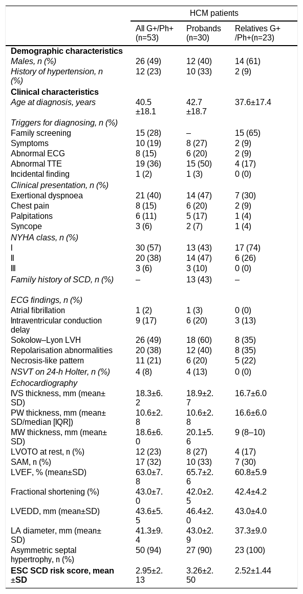
Baseline characteristics of patients with hypertrophic cardiomyopathyTo understand better the phenotypic spectrum associated with the identified MYH7 variants, we assessed the baseline clinical characteristics of the 53 affected individuals (Table 1). Of these, 49.1% were male, and the age at diagnosis was 40.5±18.1 years. The trigger for investigating subjects was the detection of increased left ventricular wall thickness on echocardiography (35.8%), family screening (28.3%), symptoms (18.9%) and abnormal ECG (15%). Most patients were in NYHA class I (74%) or II (26%).
Demographic and clinical characteristics of the included patients.
| HCM patients | |||
|---|---|---|---|
| All G+/Ph+(n=53) | Probands(n=30) | Relatives G+/Ph+(n=23) | |
| Demographic characteristics | |||
| Males, n (%) | 26 (49) | 12 (40) | 14 (61) |
| History of hypertension, n (%) | 12 (23) | 10 (33) | 2 (9) |
| Clinical characteristics | |||
| Age at diagnosis, years | 40.5±18.1 | 42.7±18.7 | 37.6±17.4 |
| Triggers for diagnosing, n (%) | |||
| Family screening | 15 (28) | – | 15 (65) |
| Symptoms | 10 (19) | 8 (27) | 2 (9) |
| Abnormal ECG | 8 (15) | 6 (20) | 2 (9) |
| Abnormal TTE | 19 (36) | 15 (50) | 4 (17) |
| Incidental finding | 1 (2) | 1 (3) | 0 (0) |
| Clinical presentation, n (%) | |||
| Exertional dyspnoea | 21 (40) | 14 (47) | 7 (30) |
| Chest pain | 8 (15) | 6 (20) | 2 (9) |
| Palpitations | 6 (11) | 5 (17) | 1 (4) |
| Syncope | 3 (6) | 2 (7) | 1 (4) |
| NYHA class, n (%) | |||
| I | 30 (57) | 13 (43) | 17 (74) |
| II | 20 (38) | 14 (47) | 6 (26) |
| III | 3 (6) | 3 (10) | 0 (0) |
| Family history of SCD, n (%) | – | 13 (43) | – |
| ECG findings, n (%) | |||
| Atrial fibrillation | 1 (2) | 1 (3) | 0 (0) |
| Intraventricular conduction delay | 9 (17) | 6 (20) | 3 (13) |
| Sokolow–Lyon LVH | 26 (49) | 18 (60) | 8 (35) |
| Repolarisation abnormalities | 20 (38) | 12 (40) | 8 (35) |
| Necrosis-like pattern | 11 (21) | 6 (20) | 5 (22) |
| NSVT on 24-h Holter, n (%) | 4 (8) | 4 (13) | 0 (0) |
| Echocardiography | |||
| IVS thickness, mm (mean±SD) | 18.3±6.2 | 18.9±2.7 | 16.7±6.0 |
| PW thickness, mm (mean±SD/median [IQR]) | 10.6±2.8 | 10.6±2.8 | 16.6±6.0 |
| MW thickness, mm (mean±SD) | 18.6±6.0 | 20.1±5.6 | 9 (8–10) |
| LVOTO at rest, n (%) | 12 (23) | 8 (27) | 4 (17) |
| SAM, n (%) | 17 (32) | 10 (33) | 7 (30) |
| LVEF, % (mean±SD) | 63.0±7.8 | 65.7±2.6 | 60.8±5.9 |
| Fractional shortening (%) | 43.0±7.0 | 42.0±2.5 | 42.4±4.2 |
| LVEDD, mm (mean±SD) | 43.6±5.5 | 46.4±2.0 | 43.0±4.0 |
| LA diameter, mm (mean±SD) | 41.3±9.4 | 43.0±2.9 | 37.3±9.0 |
| Asymmetric septal hypertrophy, n (%) | 50 (94) | 27 (90) | 23 (100) |
| ESC SCD risk score, mean±SD | 2.95±2.13 | 3.26±2.50 | 2.52±1.44 |
Values are expressed as number of patients (n) and percentage (%) for categorical variables and mean±standard deviation (mean±SD) or median (interquartile range) for continuous variables.
ECG: electrocardiogram; ESC SCD risk score: European Society of Cardiology risk score for sudden cardiac death; IVS: interventricular septum; LA: left atrium; LVEDD: left ventricular end-diastolic diameter; LVEF: left ventricular ejection fraction; LVH: left ventricular hypertrophy; LVOTO: left ventricular outflow tract obstruction; MW: maximal wall; NSVT: non-sustained ventricular tachycardia; NYHA: New York Heart Association functional class; PW: posterior wall; SAM: systolic anterior motion of the mitral valve; SCD: sudden cardiac death; TTE: transthoracic echocardiogram.
Sinus rhythm was observed in 94.3%. At diagnosis, 67.9% had an abnormal ECG: 49.1% met LVH criteria, 37.7% had ST-T abnormalities, and 20.8% had abnormal Q waves (necrosis-like pattern, mostly anteroseptal). NSVT was documented in four patients, all probands. Asymmetrical septal hypertrophy was predominant (94.3%), with mean septal thickness of 18.3±6.2 mm. At baseline, mean ESC HCM risk-SCD score was 2.92±2.13, with 15.1% of patients at intermediate or high risk. A family history of premature SCD was reported in 13 families.
No significant gender-based differences were observed regarding age at diagnosis (45.4±17.0 vs. 36.1±18.9 years, p=0.075), left atrial diameter (43.0±10.2 vs. 39.6±8.2 mm, p=0.23), LVEDD (44.9±5.2 vs. 42.2±5.7 mm, p=0.138), or septal thickness (18.2±5.7 vs. 18.5±6.7 mm, p=0.887).
Compared with affected relatives (n=23), probands (n=30) were more symptomatic (NYHA class II–III: 57% vs. 38%, p<0.001), and had a significantly larger left atrial diameter (43.0±2.9 vs. 37.3±9.0 mm, p=0.004) (Supplemental Table 1). Interventricular septal thickness (p=0.1), LVEF (p=0.148), and LVEDD (p=0.412) were not significantly different between groups.
Longitudinal follow-up data of patients with hypertrophic cardiomyopathyFunctional evolution and clinical eventsThe cohort was followed for a median of 9.5 years (interquartile range (IQR), 3.4–24.7 years; range, 0.2–46.8 years). At the last clinical evaluation, 54 patients had an HCM phenotype. Among these, 15 patients were followed for over 20 years, including 12 for more than 25 years, nine for more than 30 years, and four for over 45 years.
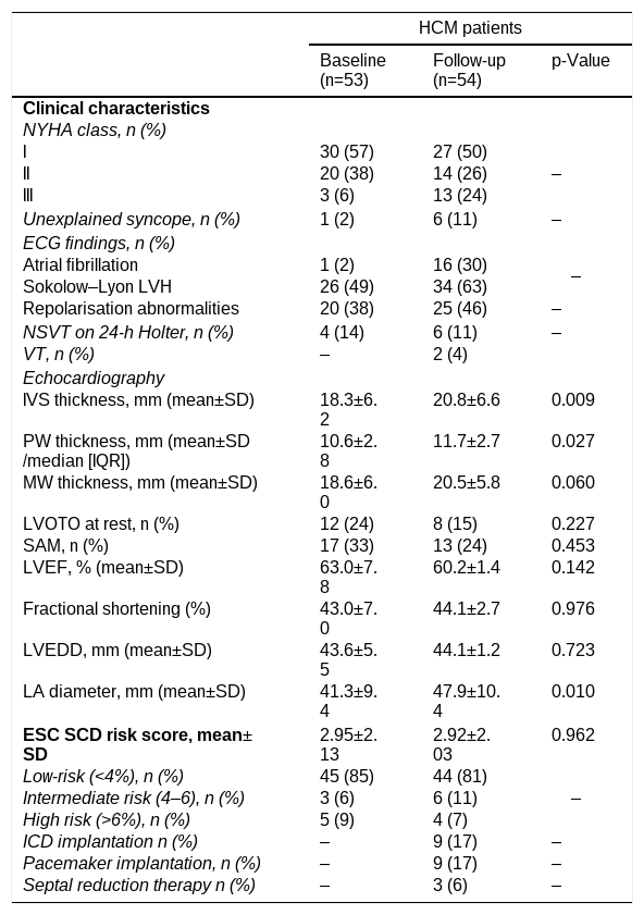
During follow-up, 14 patients (26%) experienced functional deterioration (worsening NYHA class), six developed LVOTO (no inducible gradient on exercise stress echocardiography in five tested patients), and three underwent septal reduction therapy (two alcohol septal ablations and one surgical myectomy) (Table 2).
Baseline and follow-up characteristics of the study population.
| HCM patients | |||
|---|---|---|---|
| Baseline(n=53) | Follow-up(n=54) | p-Value | |
| Clinical characteristics | |||
| NYHA class, n (%) | |||
| I | 30 (57) | 27 (50) | |
| II | 20 (38) | 14 (26) | – |
| III | 3 (6) | 13 (24) | |
| Unexplained syncope, n (%) | 1 (2) | 6 (11) | – |
| ECG findings, n (%) | |||
| Atrial fibrillation | 1 (2) | 16 (30) | – |
| Sokolow–Lyon LVH | 26 (49) | 34 (63) | |
| Repolarisation abnormalities | 20 (38) | 25 (46) | – |
| NSVT on 24-h Holter, n (%) | 4 (14) | 6 (11) | – |
| VT, n (%) | – | 2 (4) | |
| Echocardiography | |||
| IVS thickness, mm (mean±SD) | 18.3±6.2 | 20.8±6.6 | 0.009 |
| PW thickness, mm (mean±SD/median [IQR]) | 10.6±2.8 | 11.7±2.7 | 0.027 |
| MW thickness, mm (mean±SD) | 18.6±6.0 | 20.5±5.8 | 0.060 |
| LVOTO at rest, n (%) | 12 (24) | 8 (15) | 0.227 |
| SAM, n (%) | 17 (33) | 13 (24) | 0.453 |
| LVEF, % (mean±SD) | 63.0±7.8 | 60.2±1.4 | 0.142 |
| Fractional shortening (%) | 43.0±7.0 | 44.1±2.7 | 0.976 |
| LVEDD, mm (mean±SD) | 43.6±5.5 | 44.1±1.2 | 0.723 |
| LA diameter, mm (mean±SD) | 41.3±9.4 | 47.9±10.4 | 0.010 |
| ESC SCD risk score, mean±SD | 2.95±2.13 | 2.92±2.03 | 0.962 |
| Low-risk (<4%), n (%) | 45 (85) | 44 (81) | – |
| Intermediate risk (4–6), n (%) | 3 (6) | 6 (11) | |
| High risk (>6%), n (%) | 5 (9) | 4 (7) | |
| ICD implantation n (%) | – | 9 (17) | – |
| Pacemaker implantation, n (%) | – | 9 (17) | – |
| Septal reduction therapy n (%) | – | 3 (6) | – |
Values are expressed as number of patients (n) and percentage (%) for categorical variables and mean±standard deviation (mean±SD) or median (interquartile range) for continuous variables.
ECG: electrocardiogram; ESC SCD risk score: European Society of Cardiology risk score for sudden cardiac death; ICD: implantable cardioverter-defibrillator; IVS: interventricular septum; LA: left atrium; LVEDD: left ventricular end-diastolic diameter; LVEF: left ventricular ejection fraction; LVH: left ventricular hypertrophy; LVOTO: left ventricular outflow tract obstruction; MW, maximal wall; NSVT: non-sustained ventricular tachycardia; NYHA: New York Heart Association; PW: posterior wall; SAM: systolic anterior motion of the mitral valve; VT: ventricular tachycardia.
Atrial fibrillation occurred in 29.6% (n=16), with no association observed between hypertension and AF (p=0.672). Nine patients (16.7%) received ICDs for primary prevention; only one experienced appropriate shocks – an 18-year-old patient carrying the p.Arg403Gln variant, who suffered multiple appropriate shocks 5.5 years after implantation. Nine patients required a permanent pacemaker for high-grade atrioventricular (A-V) block; none had previously undergone septal reduction. Age at implantation ranged from 45 to 76 years (mean 58.4±10 years). All were low risk for SCD at the time (ESC HCM risk-SCD score 1.97±1.21%, range 1.0–3.7%), justifying the placement of pacemakers rather than ICDs.
The ESC HCM risk-SCD score remained stable during follow-up (2.92±2.03), although the proportion of patients at moderate-to-high risk increased slightly (from 15.1% to 18.5%).
Only one patient progressed to end-stage disease: A 61-year-old female (a proband carrying the p. Glu924Lys variant) diagnosed at age 26, who developed severe LVH and an obstructive phenotype. Over 22 years of follow-up she experienced progressive worsening heart failure, multiple unexplained syncopal episodes leading to ICD implantation. She later required device upgrade to CRT-D following burn-out progression. Recurrent AF further complicated the clinical course with a fast ventricular response, which was difficult to control and resulted in multiple hospitalizations. The patient was listed for heart transplantation but died while awaiting a donor.
Notably, only one carrier developed HCM during follow-up, presenting with a mild phenotype (LVH on echo) at age 39, after 25 years of clinical surveillance. The patient has remained event-free over 6.3-years of follow-up. Among carriers, one developed a left ventricular hypertrophy pattern on ECG, one developed a left anterior fascicular block, and three presented with repolarization abnormalities. No significant changes were observed in PR (mean Δ=26.8 ms, 95% CI −17.2 to 70.9, p=0.207) or QRS duration (mean Δ=91.8 ms, 95% CI −86.6 to 270.1, p=0.282) from baseline to follow-up.
Echocardiographic progressionInterventricular septal thickness increased significantly over time (18.3±6.2 to 20.8±6.6 mm, p=0.009), as did left atrial diameter (41.3±9.4 to 47.9±10.4 mm, p=0.010), while LVEDD remained stable (p=0.723). Mild left ventricular systolic dysfunction (LVEF 45–50%) was observed in five patients, while one patient developed an apical aneurysm during follow-up.
Clinical outcomes and mortalityDuring follow-up, 19 patients (35.2%) reached the composite endpoint, defined as hospitalization for heart failure, cardiovascular admissions (due to arrhythmia, acute coronary syndrome, or stroke), or all-cause death. Six patients were hospitalized for decompensated heart failure (four with preserved ejection fraction). Five patients suffered a stroke, which was fatal in three of them (annual stroke risk: 0.62%).
Sixteen patients died during follow-up (mean age at death 72.5±8.3 years, range 59–89 years), including 10 cardiovascular-related deaths (1 SCD at age 20, 6 due to heart failure, 3 due to stroke). The overall annual mortality rate was 2.0%. Five- and ten-year overall survival was 92.8% (95% confidence interval (CI): 84.1–100%) and 86.3% (95% CI: 75.5–97.1%), respectively. Only one patient had indication for heart transplantation but died while on the waiting list.
Patients who met the composite endpoint were significantly older at diagnosis (51.2±14.7 vs. 33.2±17.6 years, p<0.001) and had larger left atrial diameter (42.6±8.8 vs. 39.5±6.8 mm, p=0.048). In contrast, no significant associations were observed with cardiac biomarkers at diagnosis, including troponin T (p=0.963) and NT-proBNP (p=0.145), gender (p=0.404), family history of SCD (p=0.598), or MWT (p=0.824). Notably, none of the carriers experienced a composite endpoint event during follow-up.
Comparison between probands and relativesThe overall cohort was followed for a median of 9.5 years (IQR [interquartile range] 3.4–24.7). When stratified by subgroups, probands had a significantly shorter follow-up (median 7.17 [IQR 2.42–15.08] years) compared with relatives (17.17 [IQR 6.08–35.92] years, p=0.006). At follow-up, probands remained more symptomatic, with a higher NYHA class (2.0±0.85 vs. 1.35±0.59, p=0.005), and AF was more prevalent in this group (13 vs. 3 patients, p=0.046). Clinical and echocardiographic features – including LVEF, MWT, left atrial diameter, presence of SAM, LVOTO, NSVT, and unexplained syncope – were comparable between probands and G+/Ph+ relatives (all p>0.05).
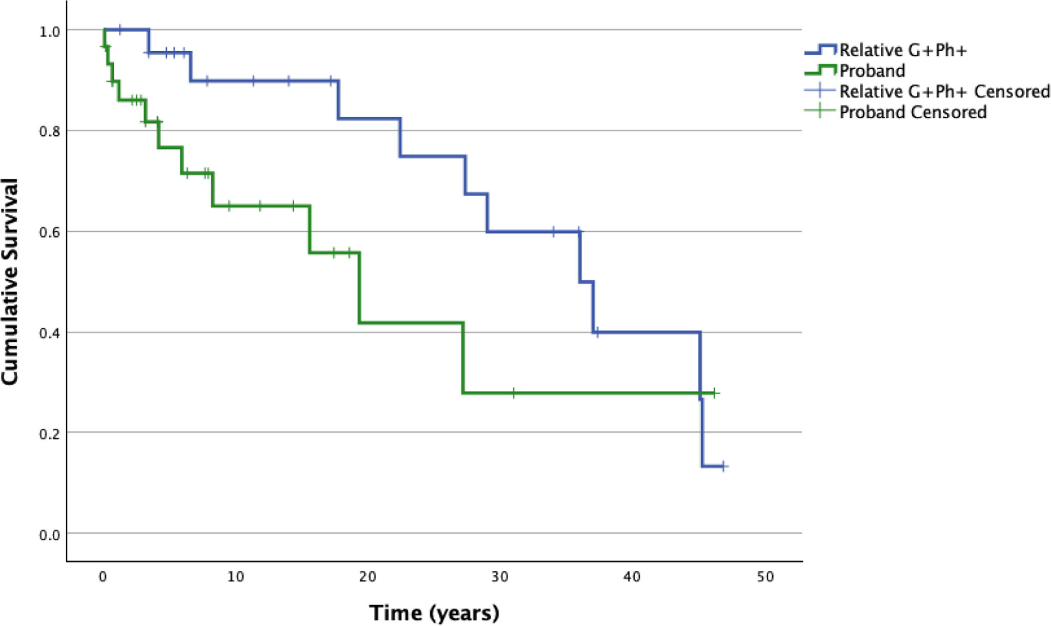
Despite the differences in follow-up, event-free survival did not differ significantly between groups, with only a non-significant trend toward worse outcomes in probands (log-rank 2.868, p=0.09; Figure 1). The presence of additional variants in probands did not significantly influence the risk of reaching the composite endpoint (log-rank 0.081, p=0.78).

Kaplan–Meier survival curves comparing event-free survival for the composite endpoint (heart failure hospitalisation, cardiovascular admissions and all-cause mortality) between probands and G+/Ph+ relatives. Although probands showed a trend toward worse event-free survival, the difference did not reach statistical significance (p=0.09; log-rank 2.868).
Thirteen distinct P/LP MYH7 variants were identified in the 30 unrelated probands (Supplemental Table 2), most of which were missense, with one small deletion. The most common variants were p.Ile263Thr (28 carriers), p.Ala797Thr (8 carriers), p.Glu1356Lys (8 carriers), and p.Arg663His (7 carriers), together accounting for 66% of all carriers. All individuals were heterozygous for the P/LP variant identified. Most variants clustered within the motor domain of MYH7 – responsible for actin binding and ATPase activity – including p.Arg403Gln, p.Arg204His, p.Arg442Cys, p.Arg719Gln, p.Arg663His, p.Ile263Thr, p.Val320Met, p.Gly741Arg. Others were located in the head–tail junction (p.Ala797Thr, p.Lys865Glu) and tail domains (p.Lys847del, p.Glu924Lys, p.Glu1356Lys), which are involved in thick filament assembly and stability (according to VarSome, accessed 21 July 2025).
An additional variant in the gene encoding for the protein ryanodine receptor 2 (RyR2), considered likely pathogenic, was identified in one family (RYR2: c.1249C>G [p.Arg417Gly]). Variants of uncertain significance or benign/likely benign were identified in six probands, involving MYBPC3, MYH6, FHL1, DOLK, NEXN, and TTN (classification according to ClinVar, accessed 21 July 2025).
Among the 77 mutation carriers, 53 fulfilled HCM diagnostic criteria, and 1 additional carrier developed phenotype during follow-up, yielding an overall penetrance of 70.1%. Among relatives identified through cascade screening, penetrance was 51.1%. Penetrance increased with age: 57.9% among carriers ≥40 years vs. 22.2% in those <40 (p<0.001) and was numerically higher in males (74.3% vs. 64.3%, p=0.437), despite a similar age at clinical evaluation (p=0.2).
Families sharing the same mutationThe four most common variants in this cohort were p.Ile263Thr (four families, 28 carriers), p.Ala797Thr (four families, eight carriers), p.Glu1356Lys (three families, eight carriers), and p.Arg663His (one family, seven carriers).
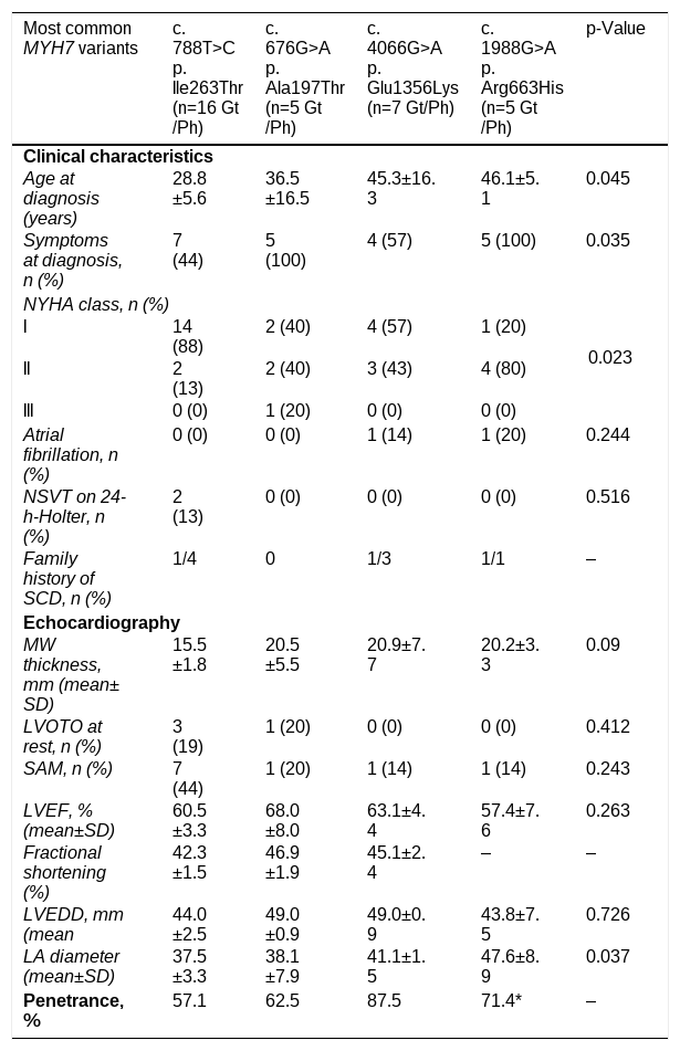
Baseline characteristics are summarized in Table 3. Given the small number of patients studied in this analysis, significant differences between variants were observed in age at diagnosis (p=0.045), the proportion of symptomatic individuals (p=0.035), NYHA class at presentation (p=0.023), and left atrial diameter (p=0.037).
Baseline clinical and echocardiographic characteristics of HCM patients carrying the four most common MYH7 mutations.
| Most common MYH7 variants | c.788T>C p.Ile263Thr(n=16 Gt/Ph) | c.676G>A p.Ala197Thr(n=5 Gt/Ph) | c.4066G>A p.Glu1356Lys(n=7 Gt/Ph) | c.1988G>A p.Arg663His(n=5 Gt/Ph) | p-Value |
|---|---|---|---|---|---|
| Clinical characteristics | |||||
| Age at diagnosis (years) | 28.8±5.6 | 36.5±16.5 | 45.3±16.3 | 46.1±5.1 | 0.045 |
| Symptoms at diagnosis, n (%) | 7 (44) | 5 (100) | 4 (57) | 5 (100) | 0.035 |
| NYHA class, n (%) | |||||
| I | 14 (88) | 2 (40) | 4 (57) | 1 (20) | 0.023 |
| II | 2 (13) | 2 (40) | 3 (43) | 4 (80) | |
| III | 0 (0) | 1 (20) | 0 (0) | 0 (0) | |
| Atrial fibrillation, n (%) | 0 (0) | 0 (0) | 1 (14) | 1 (20) | 0.244 |
| NSVT on 24-h-Holter, n (%) | 2 (13) | 0 (0) | 0 (0) | 0 (0) | 0.516 |
| Family history of SCD, n (%) | 1/4 | 0 | 1/3 | 1/1 | – |
| Echocardiography | |||||
| MW thickness, mm (mean±SD) | 15.5±1.8 | 20.5±5.5 | 20.9±7.7 | 20.2±3.3 | 0.09 |
| LVOTO at rest, n (%) | 3 (19) | 1 (20) | 0 (0) | 0 (0) | 0.412 |
| SAM, n (%) | 7 (44) | 1 (20) | 1 (14) | 1 (14) | 0.243 |
| LVEF, % (mean±SD) | 60.5±3.3 | 68.0±8.0 | 63.1±4.4 | 57.4±7.6 | 0.263 |
| Fractional shortening (%) | 42.3±1.5 | 46.9±1.9 | 45.1±2.4 | – | – |
| LVEDD, mm (mean | 44.0±2.5 | 49.0±0.9 | 49.0±0.9 | 43.8±7.5 | 0.726 |
| LA diameter (mean±SD) | 37.5±3.3 | 38.1±7.9 | 41.1±1.5 | 47.6±8.9 | 0.037 |
| Penetrance, % | 57.1 | 62.5 | 87.5 | 71.4* | – |
Values are expressed as number of patients (n) and percentage (%) for categorical variables and mean±standard deviation (mean±SD) or median (interquartile range) for continuous variables.
LA: left atrium; LVEDD: left ventricular end-diastolic diameter; LVEF: left ventricular ejection fraction; LVOTO: left ventricular outflow tract obstruction; MWT: maximal wall thickness; NSVT: non-sustained ventricular tachycardia; NYHA class: New York Heart Association; SAM: systolic anterior motion; SCD: sudden cardiac death.
Post-hoc analysis revealed that p.Arg663His carriers were diagnosed at a significantly older age compared to those with p.Ile263Thr (p=0.039) and had larger left atrial diameters (p=0.044). Notably, all carriers of p.Arg663His and p.Ala797Thr were symptomatic at diagnosis. A family history of SCD was reported in probands carrying the p.Arg663His, p.Ile263Thr, and p.Glu1356Lys variants.
Over a median follow-up of 14.0 years (IQR 4.75–29.0), phenotypic penetrance was 64.7%. Penetrance was highest in carriers of the p.Glu1356Lys (87.5%) and p.Arg663His (85.7%) variants, and lower in those with p.Ala797Thr (62.5%) and p.Ile263Thr (57.1%) (Table 3).
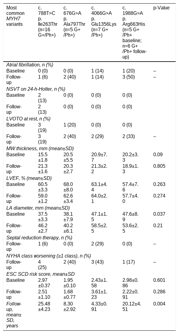
During follow-up, only one previously asymptomatic carrier, with the p.Arg663His variant, developed the phenotype. Overall, five patients developed LVOTO (nine affected at last visit), five developed AF (seven affected overall), 10 patients experienced NYHA class worsening, and 3 patients underwent septal reduction therapy, with no differences between mutations (Table 4). NSVT occurred in two p.Ile263Thr carriers, both receiving ICDs (p=0.687). ESC HCM risk-SCD scores were similar across variants both at baseline (p=0.601) and at last follow-up (p=0.286).
Comparison of baseline and follow-up clinical and echocardiographic characteristics among HCM patients carrying the four most prevalent MYH7 variants.
| Most common MYH7 variants | c.788T>C p.Ile263Thr(n=16 G+/Ph+) | c.676G>A p.Ala797Thr(n=5 G+/Ph+) | c.4066G>A p.Glu1356Lys(n=7 G+/Ph+) | c.1988G>A p.Arg663His(n=5 G+/Ph+ baseline; n=6 G+/Ph+ follow-up) | p-Value |
|---|---|---|---|---|---|
| Atrial fibrillation, n (%) | |||||
| Baseline | 0 (0) | 0 (0) | 1 (14) | 1 (20) | – |
| Follow-up | 1 (6) | 2 (40) | 1 (14) | 3 (50) | – |
| NSVT on 24-h-Holter, n (%) | |||||
| Baseline | 2 (13) | 0 (0) | 0 (0) | 0 (0) | |
| Follow-up | 2 (13) | 0 (0) | 0 (0) | 0 (0) | |
| LVOTO at rest, n (%) | |||||
| Baseline | 3 (19) | 1 (20) | 0 (0) | 0 (0) | |
| Follow-up | 3 (19) | 2 (40) | 2 (29) | 2 (33) | – |
| MW thickness, mm (mean±SD) | |||||
| Baseline | 15.5±1.8 | 20.5±5.5 | 20.9±7.7 | 20.2±3.3 | 0.09 |
| Follow-up | 21.3±1.6 | 20.3±2.7 | 21.3±2.2 | 18.9±1.3 | 0.805 |
| LVEF, % (mean±SD) | |||||
| Baseline | 60.5±3.3 | 68.0±8.0 | 63.1±4.4 | 57.4±7.6 | 0.263 |
| Follow-up | 59.0±1.2 | 62.6±3.4 | 64.0±2.1 | 57.7±4.0 | 0.274 |
| LA diameter, mm (mean±SD) | |||||
| Baseline | 37.5±3.3 | 38.1±7.9 | 47.1±1.5 | 47.6±8.9 | 0.037 |
| Follow-up | 46.2±2.7 | 40.2±6.1 | 58.5±2.5 | 53.6±2.5 | 0.21 |
| Septal reduction therapy, n (%) | |||||
| Follow-up | 1 (6) | 0 (0) | 2 (29) | 0 (0) | – |
| NYHA class worsening (≥1 class), n (%) | |||||
| Follow-up | 4 (25) | 2 (40) | 3 (43) | 1 (17) | – |
| ESC SCD risk score, mean±SD | |||||
| Baseline | 2.97±0.37 | 1.95±0.10 | 2.43±1.58 | 2.98±0.86 | 0.601 |
| Follow-up | 2.51±1.10 | 1.68±0.77 | 3.61±1.23 | 2.22±0.91 | 0.286 |
| Follow-up, mean±SD, years | 25.48±4.23 | 8.30±2.92 | 4.33±0.91 | 20.12±4.51 | 0.004 |
Values are expressed as number of patients (n) and percentage (%) for categorical variables and mean±standard deviation (mean±SD) or median (interquartile range) for continuous variables.
ESC SCD risk score: European Society of Cardiology sudden cardiac death risk score; LA: left atrium; LVEF: left ventricular ejection fraction; LVOTO: left ventricular outflow tract obstruction; MWT: maximal wall thickness; NYHA: New York Heart Association functional class; NSVT: non-sustained ventricular tachycardia.
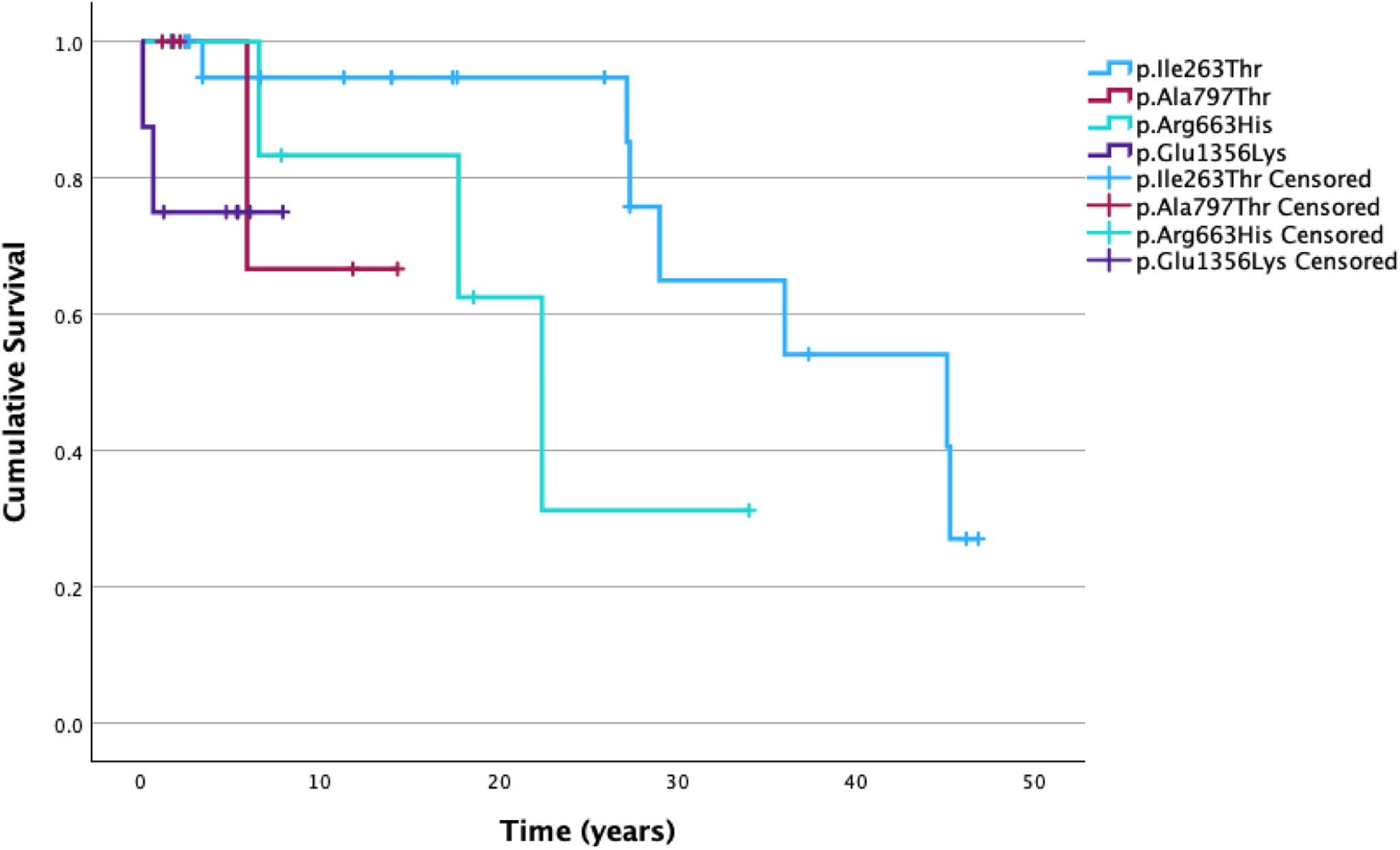
Regarding disease progression, no significant differences were observed in left atrial diameter (p=0.21), LVEF (ΔLVEF, p=0.86), or MWT (ΔMWT, p=0.72) across variants. The composite endpoint of heart failure hospitalization, cardiovascular hospitalization, or all-cause mortality occurred similarly across variant groups (log-rank 7.3, p=0.06; Figure 2), despite differences in follow-up duration (p=0.004).

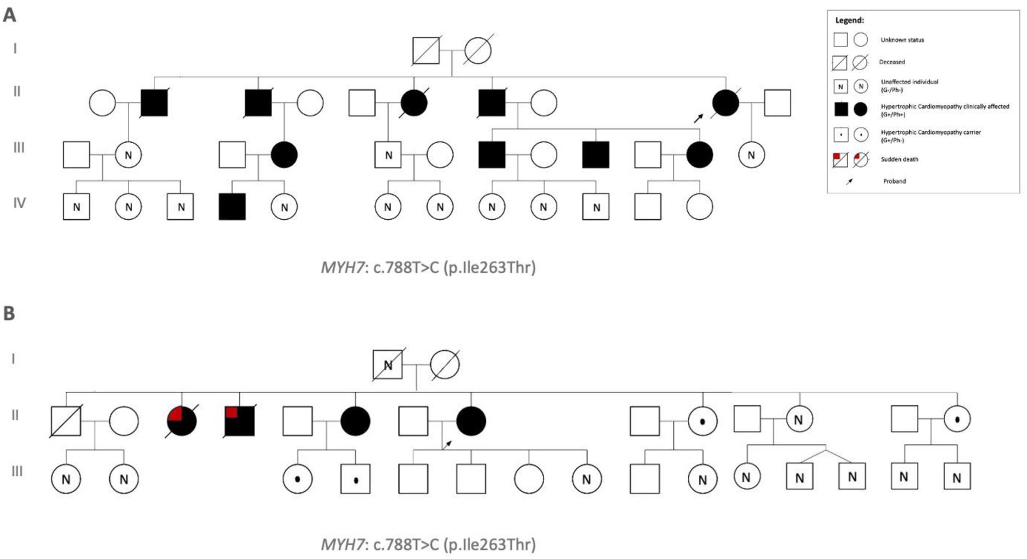
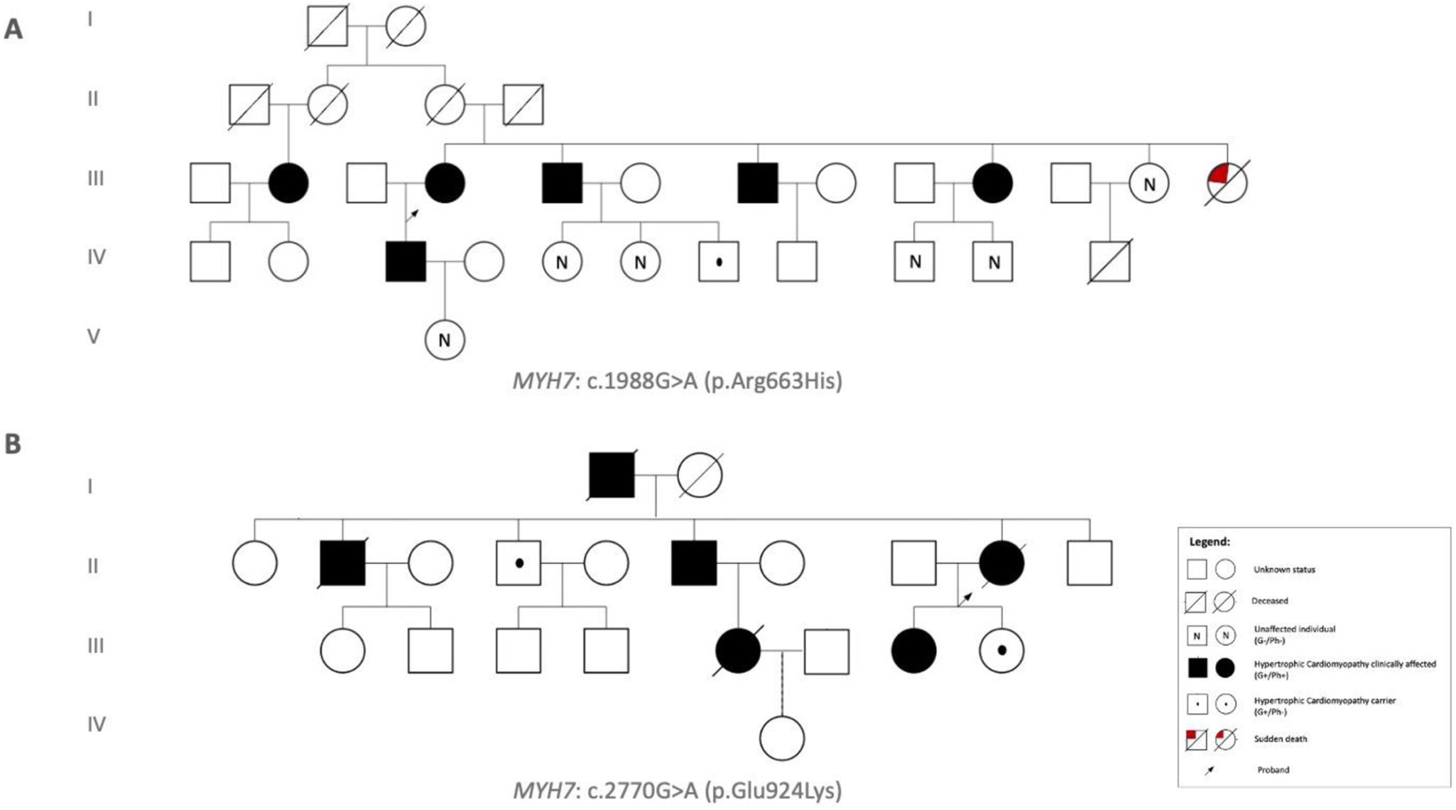
However, phenotypic expression was highly variable between families (Figures 3 and 4), and even among those carrying the same mutation, penetrance and clinical course differed.


In the four families with the p.Ile263Thr variant, 16 out of the 28 carriers met HCM criteria (penetrance 57.1%). But one family showed complete penetrance while in the other families the penetrance ranged from 33% to 50%. Age at diagnosis varied widely, from 17 years in a patient with a murmur that led to the prohibition of sports and initiation of diagnostic workup, to 42 years in another patient diagnosed after ECG repolarization abnormalities. One large family (19 individuals studied) exhibited moderate to severe LVH (MWT 18.3±2 mm), with only one individual experiencing AF, but two cases of SCD under 30 years of age and one death from heart failure occurred. Another family, during a mean follow-up of 28.4±15.5 years (range 3–46), showed more severe LVH (MWT 23±6 mm), but only the proband had cardiovascular death (hemorrhagic stroke), with no other patients experiencing significant symptoms, had indication for ICD implantation, or suffered SCD. In the other two families, only the proband is phenotypically affected.
Among p.Ala797Thr carriers (four families, seven carriers), a proband was diagnosed at age 53 with moderate LVH (MWT 17.3 mm) and LVOTO on echocardiogram, remained asymptomatic on beta-blocker therapy, with no arrhythmias or heart failure events; his three children remained asymptomatic carries after 6 years of follow-up (at age 26, 26, 30). In contrast, another 19-year-old proband developed severe LVH (MWT 31 mm) and NYHA III symptoms. Only one patient developed AF during a median follow-up of 11.8 years (IQR 1.25–13.6). No cases of SCD, ventricular tachycardia, stroke, or heart failure hospitalization occurred in any of these families. Penetrance ranged from 25% to 100%.
In p.Glu1356Lys carriers (three families, seven carriers), phenotypic penetrance ranged from 75% to 100%. One proband was diagnosed at 58 years by echocardiogram, after receiving a pacemaker at 48 years of age; his niece identified through family screening, had severe septal hypertrophy (MWT 35 mm), underwent myectomy at 17 years of age, and received an ICD for primary prevention (ESC HCM risk-SCD score of 5.6%). Among three other carriers, one had HCM criteria: a 49-year-old male with mild septal hypertrophy, that had severe left atrial dilation (58 mm) and experienced one episode of NSVT. In another family, the proband presented at 55 years of age with syncope and mild to moderate LVH; he underwent septal alcohol ablation during follow-up. Despite a persistent significant subaortic gradient, she declined surgical myectomy and died 2 years later. In the third family, the proband was diagnosed incidentally at 65 years of age, later developed AF, and also received a pacemaker due to A-V block.
The p.Arg663His family (seven carriers) showed high penetrance over long-term follow-up (85.7%) with predominantly late-onset phenotypes. The proband, diagnosed at 45 (MWT of 26 mm), developed AF but remained asymptomatic into his 80s. His son developed mild hypertrophy (MWT 13.9 mm) after 26 years and remained asymptomatic. Four additional relatives met HCM criteria, including two who also have AF, one that suffered a heart failure hospitalization, and two patients with mild, asymptomatic disease. A family history of SCD was reported.
DiscussionIn this single-center cohort of individuals carrying P/LP MYH7 variants, we describe the natural history, phenotypic spectrum, and long-term clinical progression of HCM. To our knowledge, this is one of the longest observational studies of genetically confirmed MYH7 variant carriers, with comprehensive phenotypic characterization and with 27% of patients being followed up over 20 years, extending up to more than 40 years in some patients.
The major findings were: (1) very heterogeneous clinical disease expression and not clearly related to the specific variants; (2) incomplete and variable disease penetrance, even among families harboring the same mutation; (3) probands exhibited a more severe phenotype at baseline but did not suffer worse outcomes during follow-up; (4) AF occurred in approximately one-third of patients; (5) the risk of SCD was clinically relevant; (6) over long-term serial assessments, symptoms were common, with more than one-third of patients reaching a composite endpoint of heart failure hospitalization, cardiovascular admissions, or all-cause death.
In 30 unrelated families, 13 distinct P/LP mutations were identified, highlighting the marked genetic heterogeneity of the disease. This heterogeneity is associated, at least in part, with variability in clinical manifestations, which makes it challenging to establish clear genotype–phenotype correlations.19MYH7-associated HCM has been characterized by early disease onset13 and generally more severe phenotypes compared with MYBPC3 mutations.13,14
The cohort's median age at onset was consistent with prior reports, typically in the early 40s,12 with a wide range from childhood (eight years old) to the seventh decade of life, with a higher penetrance in males.20 Experimental data suggest that estrogen may modulate hypertrophic pathways epigenetically, potentially delaying disease manifestation in females.21 Nevertheless, clinical severity and event rates were similar between genders. This is supported by a multicenter study by Olivotto et al. (969 HCM patients), in which male predominance (59%) did not translate into differences in mortality or sudden cardiac death.22
A recent meta-analysis including 307 relatives carrying MYH7 variants reported an overall disease penetrance of 64%, with wide variability (40–100%) across studies.23 In this cohort, penetrance was 70% and increased progressively with age – reinforcing the importance of lifelong clinical surveillance in genotype-positive individuals, as HCM may have late penetrance.24 Notably, despite the extended follow-up duration, only one patient developed a clinical phenotype after more than 25 years of surveillance, contrasting with previous reports indicating phenotypic conversion rates of up to 23% over approximately nine years of follow-up.23
It is now well recognized that HCM is frequently a benign disease.24 In this cohort, baseline phenotypes were generally mild, with moderate LVH, mild left atrial enlargement, and low ESC HCM risk-SCD score. Nonetheless, family history of SCD was present in 13 families. As expected, probands had more severe disease (and accounted for all cases of NSVT), likely due to earlier detection in relatives through cascade screening and referral bias in symptomatic probands. Despite this, outcomes did not significantly differ between probands and relatives. Over time, most patients progressed to moderate-to-severe LVH, moderate left atrial enlargement, consistent with previous reports,12,13,25 and worsening NYHA class in ∼25%. Although average ESC SCD risk scores remained stable, more patients transitioned to intermediate/high-risk categories, underlining the need for serial risk reassessment.
Decisions regarding ICD implantation in this study population are based on established risk scores and clinical judgment. The current risk score relies on maximal interventricular septal thickness as a key parameter. However, Olivotto et al.26 have shown that left ventricular mass indexed to body surface area is a more sensitive predictor of HCM-related death than MWT. Moreover, MWT – a cornerstone metric for diagnosis and risk stratification – exhibits significant interobserver variability across imaging modalities. In a large expert-based study, echocardiographic (±20%) and cardiac magnetic resonance (CMR) (±11%) measurements showed poor correlation (r=0.56), with discrepancies affecting ICD eligibility in approximately 14% of patients.27 In this cohort, among nine patients who received an ICD for primary prevention, only one experienced appropriate shock during follow-up, highlighting the challenge of accurately identifying patients truly at risk of life-threatening arrhythmia.
Atrial fibrillation emerged as a major burden in the MYH7 cohort, affecting approximately one-third of patients – higher than the 19–24% reported in previous HCM series.12,25,28 In the study by Lee et al., which included 1040 HCM patients (mean age 40.1±15.2 years, 59.2% male) followed over 7.2 years, AF occurred in 19%.28 Prior evidence suggests that patients with MYH7 variants were more likely to develop AF compared to those carrying MYBPC3 or thin filament variants (log-rank, p=0.016), especially in the presence of larger left atria, higher resting LVOT gradient, and greater MWT.12 This highlights the importance of close surveillance in MYH7-HCM patients, enabling early detection of AF and timely initiation of appropriate management strategies, including prompt oral anticoagulation irrespective of CHA2DS2-VASc score.29
In contrast, the burden of ventricular arrhythmias in this population was lower than previously reported. Sedaghat-Hamedani et al.12 observed ventricular tachycardia in 33% and conduction abnormalities in 31% of MY7H7 carriers. Among patients in this cohort, NSVT was detected in 11%, sustained ventricular tachycardia in 3.7%, while 17% underwent ICD implantation for primary prevention and 17% required a pacemaker. These differences may be explained by variation in sample size, by different mutations that may have distinct functional consequences, and by different follow-up protocols.
Despite a relatively benign initial presentation and contemporary advances in the management of HCM – which is now associated with mortality approaching that of the general population29 – adverse outcomes were notable: 11% experienced heart failure hospitalization, 9% had a stroke, and 10 cardiovascular deaths occurred, including one SCD. The risk of SCD aligns with published rates of 4–12%.12,30 Overall annual mortality rate was 2.0%, with older age at diagnosis and larger left atrial size predicting a worse prognosis. Heart transplantation is a relevant endpoint in sarcomeric HCM. Previous studies reported a 7.7% transplant rate in MYH7-associated HCM.23 In this cohort, only one patient was referred for heart transplant but did not undergo the procedure.
During long-term follow-up, the only individual who developed a phenotype also carried a benign/likely benign MYBPC3 mutation. In this cohort, most double-variant carriers harbored variants of uncertain significance, which may explain why multiple variants were not associated with a more severe phenotype or adverse outcomes. This contrasts with previous reports suggesting that 5–7% of HCM patients carry two or more sarcomeric mutations,31 and that compound heterozygosity is associated with more severe disease and higher rates of cardiovascular death.31,32 However, the problem of variant classification and the need for re-classification at least every two years33 is a genuine issue, namely for variants of uncertain significance.34
Despite broadly similar outcomes across the four most prevalent variants, the penetrance of each variant differed, with some distinct phenotypic features that may carry clinical relevance.
In particular, carriers of p.Arg663His, exhibited all these three characteristics: they were consistently symptomatic, diagnosed at a later age and had a larger left atrium. Phenotypic penetrance for this variant was 86%, higher than previously described (50–72%).35 For the p.Ile263Thr variant, external data are scarce; while this variant has been reported in Portuguese,36,37 French31 and Brazilian25 cohorts, detailed phenotypic characterization is lacking. In this cohort, two carriers of p.Ile263Thr underwent ICD implantation for primary prevention.
Both of these variants are located in the motor domain and appear to be associated with a slightly more severe clinical profile, but further studies are needed to confirm this observation. In a small cohort, it is difficult to detect statistically significant differences that would allow robust delineation of a genotype–phenotype risk profile, and no such associations were identified.
The p.Ala797Thr variant showed broad variability in age at onset and modest phenotypes, with no severe outcomes observed in this cohort. This variant, previously described as a founder mutation in South African Afrikaners,37 has been associated with moderate LVH, favorable long-term prognosis and normal life span. Nonetheless, disease expression can vary substantially even within the same family – as illustrated by one family in the study cohort37 in which three cases of SCD occurred. In that cohort, disease penetrance was 61%, similar to our findings.
The p.Glu1356Lys variant has previously been linked to arrhythmias, ICD use, and septal reduction therapy, with a later diagnosis age (55–65 years) among probands.38 Within the study cohort, it was likewise associated with later diagnosis and demonstrated the highest phenotypic penetrance (87.5%). Overall, the small number of individuals per variant limits the ability to draw definitive genotype–phenotype correlations, highlighting the need for larger studies to better characterize variant-specific disease expression.
This study has several limitations. It is a single-center cohort of predominantly White European patients, which may limit generalizability. Although follow-up was long, the retrospective nature of data collection may introduce bias. However prospective long longitudinal studies in HCM would demand a follow up over decades with an analysis of what happened during the patients’ lifetime after diagnosis. It was in fact what our center did: to follow HCM patients during their lifetime. CMR data were not included, as their availability was limited by the historical nature of the cohort, while echocardiography was consistently performed at all follow-up visits. Although CMR is not recommended for routine screening of HCM in asymptomatic genetic carriers, we acknowledge that its absence represents a limitation, as subtle phenotypic features could theoretically have been missed in some patients. The small number of clinical events and small numbers within each variant group (private mutations), limit direct comparisons and reduce statistical power.
ConclusionsThe spectrum of clinical disease expression observed in individuals and families with MYH7 mutations is broad and variable. In this cohort, which included 13 different pathogenic/likely pathogenic mutations in 30 unrelated families with HCM, the majority of patients with the disease presented with a benign phenotype over a long follow-up period. However, the risk of SCD existed in 17% of patients, requiring preventive ICD implantation. Additionally, more than one-third of patients experienced a composite endpoint including heart failure hospitalization, cardiovascular admission (for arrhythmia, acute coronary syndrome, or stroke), or all-cause mortality.
Despite the small number of individuals studied, analysis of the four most common variants in this cohort revealed differing penetrance but similar clinical outcomes, highlighting the difficulty of defining a clinical profile for each variant and illustrating once more why the absence of genotype in current HCM risk models.
Clinicians should therefore use available genotype–phenotype data to inform counselling of at-risk family members, while recognizing the substantial variability in clinical expression and disease progression.
Ethical approvalThis study was conducted in accordance with the principles of the Declaration of Helsinki. As this is a retrospective observational study without intervention, formal approval from the ethics committee was not required (registration number not applicable).
FundingThis research received no specific grant from any funding agency in the public, commercial, or not-for-profit sectors.
Conflicts of interestThe authors have no conflicts of interest related to the content of this study.
The following are the supplementary data to this article: