
Sudden cardiac death (SCD) risk stratification in non-ischemic cardiomyopathy (NICM) remains challenging. The recently proposed Derivate Risk Score 2.0 (DRS 2.0) seeks to enhance risk prediction using three variables: sex, cardiac magnetic resonance-derived left ventricular ejection fraction (CMR-LVEF), and the presence of midwall late gadolinium enhancement (LGE). The aim of this study was to assess the accuracy of the DRS 2.0 for predicting Major arrhythmic events (MAACE) in a Portuguese NICM cohort.
MethodsPatients with the clinical diagnosis of NICM who underwent CMR for clinical purposes were included in this single-center retrospective study. Patients with obstructive coronary artery disease, infiltrative, inflammatory or hypertrophic cardiomyopathy, or significant valvular or congenital heart disease were excluded. The DRS 2.0 score was calculated for each patient based on male sex, CMR-derived LVEF and on the presence and pattern of midwall LGE as reported. The primary endpoint was major arrhythmic cardiac events (MAACE), defined as SCD, sustained ventricular tachycardia (VT), or appropriate ICD therapy (shock or ATP). Follow-up was performed using clinical records and device interrogations.
ResultsA total of 385 patients were enrolled (age 62.5 years (±14.9); 63% male, median CMR-LVEF 37% (27–43), midwall LGE present 51.4%). One hundred two patients had an ICD (26.5%), 5 had an ICD previous to CMR and the remaining 97 patients received one during follow up. The median DRS2.0 score was 6.0 (IQR 4.0–9.0). Risk distribution according to the DRS 2.0 was 20% low risk (n=76), 53% intermediate risk (n=203), and 28% high risk (n=106). During a mean follow-up of 3.0±1.4 years, 26 MAACE (6.8%) occurred, including 7 SCD, 9 sustained VT, and 10 appropriate ICD therapies. The annualized MAACE rates were 0.4%/year in low risk, 1.0%/year in intermediate-risk, and 6.0%/year in the high-risk patients. Patients classified as high risk by DRS 2.0 represented 28% of the cohort yet accounted for 73% of all MAACE (HR 8.00, 95% CI 3.36–19.04, p<0.001). The DRS 2.0 showed good discrimination (C statistic 0.76, 95% CI: 0.66–0.85, p<0.001). Calibration was excellent, as indicated by a non-significant Hosmer–Lemeshow test (χ2=0.802, p=0.371).
ConclusionsDRS 2.0 showed robust discrimination and calibration in our Portuguese NICM cohort, effectively identifying a small subgroup that accounted for most arrhythmic events. This score appears applicable in our population and may assist in refining SCD risk stratification and management.
A estratificação do risco de morte súbita cardíaca (MSC) na miocardiopatia não isquémica (MNI) permanece um desafio. O recentemente proposto DERIVATE Risk Score 2.0 (DRS 2.0) procura melhorar a previsão de risco utilizando três variáveis: sexo masculino, fração de ejeção do ventrículo esquerdo medida por ressonância magnética cardíaca (FEVE-RMC) e a presença de realce tardio com gadolínio (RTG) médio-mural. O objetivo deste estudo foi avaliar o desempenho do DRS 2.0 na previsão de eventos arrítmicos major (MAACE) numa coorte portuguesa de MNI.
MétodosForam incluídos neste estudo retrospetivo unicêntrico doentes com diagnóstico clínico de MNI submetidos a RMC por indicação clínica. Foram excluídos doentes com doença coronária obstrutiva, miocardiopatia infiltrativa, inflamatória ou hipertrófica, ou doença valvular ou congénita significativa. A pontuação DRS 2.0 foi calculada para cada doente com base no sexo masculino, FEVE-RMC e na presença e padrão de RTG médio-mural, conforme reportado. O endpoint primário foi a ocorrência de eventos arrítmicos major (MAACE), definidos como MSC, taquicardia ventricular (TV) sustentada ou terapêutica apropriada de CDI (choque ou ATP). O seguimento foi realizado através de registos clínicos e interrogação de dispositivos.
ResultadosForam incluídos um total de 385 doentes (idade 62,5 ± 14,9 anos; 63% sexo masculino, FEVE-RMC mediana 37% (27-43), presença de RTG médio-mural em 51,4%). 102 doentes tinham CDI (26,5%), 5 dos quais já tinham o dispositivo antes da RMC e os restantes 97 receberam-no durante o seguimento. A pontuação mediana do DRS 2.0 foi de 6,0 (IIQ 4,0–9,0). A distribuição de risco de acordo com o DRS 2.0 foi de 20% de baixo risco (n=76), 53% de risco intermédio (n=203) e 28% de alto risco (n=106). Durante um seguimento médio de 3,0 ± 1,4 anos, ocorreram 26 MAACE (6,8%), incluindo 7 MSC, 9 TV sustentadas e 10 terapêuticas apropriadas de CDI. As taxas anualizadas de MAACE foram de 0,4%/ano no baixo risco, 1,0%/ano no risco intermédio e 6,0%/ano nos doentes de alto risco. Os doentes classificados como de alto risco pelo DRS 2.0 representaram 28% da coorte, mas foram responsáveis por 73% de todos os MAACE (HR 8,00, IC 95% 3,36-19,04, p<0,001). O DRS 2.0 apresentou uma boa discriminação (C statistics 0,76, IC 95%: 0,66-0,85, p<0,001). A calibração foi excelente, conforme indicado pelo teste de Hosmer-Lemeshow não significativo (Xn2=0,802, p=0,371).
ConclusõesO DRS 2.0 demonstrou uma boa discriminação e excelente calibração na nossa coorte portuguesa de MNI, identificando eficazmente um pequeno subgrupo que concentrou a maioria dos eventos arrítmicos. Este score parece ser aplicável na nossa população e poderá ser uma ferramenta útil na estratificação do risco de morte súbita em doente com MNI.

Sudden cardiac death (SCD) remains one of the most devastating outcomes of non-ischemic cardiomyopathy (NICM). Although recent data suggest a decline in SCD incidence – likely reflecting advances in pharmacological therapy and heart failure management – accurately identifying patients at highest risk for malignant ventricular arrhythmias remains a major clinical challenge.1–3 The unpredictable occurrence of these events, often in patients who do not meet conventional high-risk criteria, highlights the need for improved risk stratification strategies.4
For decades, primary prevention implantable cardioverter-defibrillator (ICD) therapy has been guided almost exclusively by a left ventricular ejection fraction (LVEF) threshold of <35%, based largely on landmark clinical trials.5,6 However, this approach has important limitations. While reduced LVEF reflects heart failure severity, it lacks both sensitivity and specificity for predicting arrhythmic risk in patients with NICM.7,8
Cardiac magnetic resonance (CMR) imaging has emerged as a powerful tool to address these limitations. Beyond accurate LVEF quantification, CMR enables the detection of myocardial fibrosis using late gadolinium enhancement (LGE). In particular, midwall LGE has been identified as a strong arrhythmogenic substrate, independent of systolic function.9 Alongside imaging, genetic information has also been important in the evaluation of NICM, as specific genotypes are known to confer a high risk of arrhythmic events.10
The DERIVATE Risk Score 2.0 (DRS 2.0) was recently introduced as a multidimensional risk prediction model. By combining male sex, CMR-derived LVEF, and the presence and pattern of midwall LGE, the score demonstrated high diagnostic accuracy and prognostic value in its derivation and validation cohorts.4
However, its performance across different geographic regions and clinical settings remains uncertain. Given potential variations in genetic background, clinical practice, and referral patterns, external validation in diverse populations is essential to support broader clinical implementation.
ObjectivesThis study aimed to evaluate the performance, discrimination, and calibration of the DRS 2.0 for predicting major arrhythmic events in a contemporary Portuguese cohort of patients with NICM.
MethodsStudy designThis was a single-center observational study conducted at Hospital de Santa Cruz, Lisbon, Portugal. Consecutive cardiac magnetic resonance (CMR) examinations performed for clinical indications between 2019 and 2024 were analysed. Eligible patients were aged ≥18 years, had chronic heart failure according to European Society of Cardiology Task Force criteria, and a CMR-derived left ventricular ejection fraction (LVEF) <50%.
Patients with obstructive coronary artery disease, infiltrative, inflammatory, or hypertrophic cardiomyopathies, as well as those with significant valvular or congenital heart disease, were excluded. Baseline characteristics, including demographic data, clinical information, and medical and device therapy, were defined at the time of the index CMR and obtained from medical records corresponding to the clinical evaluation closest to the MRI date. Genetic data was not gathered since it was not included as variables in the calculation of the DRS 2.0.
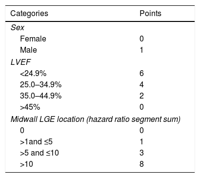
The DERIVATE Risk Score 2.0 (DRS 2.0) was calculated for each patient using male sex, CMR-derived LVEF, and the presence and pattern of midwall late gadolinium enhancement (LGE), as detailed in Table 1. Patients were stratified into three risk categories: low (≤3 points), intermediate (4–9 points), and high (>9 points).
The primary endpoint was major arrhythmic cardiac events (MAACE), defined as sudden cardiac death, sustained ventricular tachycardia (>30 seconds), or appropriate implantable cardioverter-defibrillator therapy (shock or antitachycardia pacing).1 Clinical events were analyzed from the date of the CMR until October 31, 2025. Follow-up data were obtained from clinical records and device interrogations.
Cardiac magnetic resonanceAll cardiac magnetic resonance (CMR) studies were performed using a 1.5-Tesla scanner (Magnetom Avanto; Siemens Medical Solutions, Erlangen, Germany) following a standardized institutional protocol. Image post-processing and quantitative analysis were conducted with dedicated software (Circle Cardiovascular Imaging, CVI version 5.6.4, Calgary, Canada). Left ventricular (LV) volumes, mass, and ejection fraction were measured using standard volumetric techniques, based on endocardial and epicardial contour delineation in contiguous short-axis cine images acquired at end-diastole and end-systole.
Late gadolinium enhancement (LGE) images were acquired approximately 10 minutes after intravenous administration of 0.2 mmol/kg of gadolinium-based contrast, using breath-hold segmented inversion-recovery or phase-sensitive inversion-recovery sequences. The LV short-axis LGE stack was visually assessed for the presence and pattern of LGE.
Statistical analysisStatistical analyses were performed using SPSS version 30 (SPSS Inc., Chicago, IL, USA) and JASP version 0.95.4 (JASP Team, Amsterdam, The Netherlands). Categorical variables were expressed as frequencies and percentages, while continuous variables were reported as mean±standard deviation or median with interquartile range, as appropriate. Comparisons between categorical variables were performed using the chi-square test or Fisher's exact test. Continuous variables were compared using the Student's t-test or the Mann–Whitney U test, depending on data distribution. Multivariable analysis was not performed due to the limited number of events.
Cumulative incidence of major arrhythmic cardiac events (MAACE) during follow-up was estimated using the Kaplan–Meier method, with differences in event-free survival between risk groups assessed using the log-rank (Mantel–Cox) test. The performance of the DERIVATE Risk Score 2.0 (DRS 2.0) was evaluated using both predictive and discriminative metrics. Discrimination was assessed by receiver operating characteristic (ROC) curve analysis, with calculation of the area under the curve (AUC), and by Harrell's C-index to account for censored time-to-event data. Model calibration was evaluated using the Hosmer–Lemeshow goodness-of-fit test, comparing observed and expected MAACE rates. All tests were two-sided, and a p-value <0.05 was considered statistically significant.
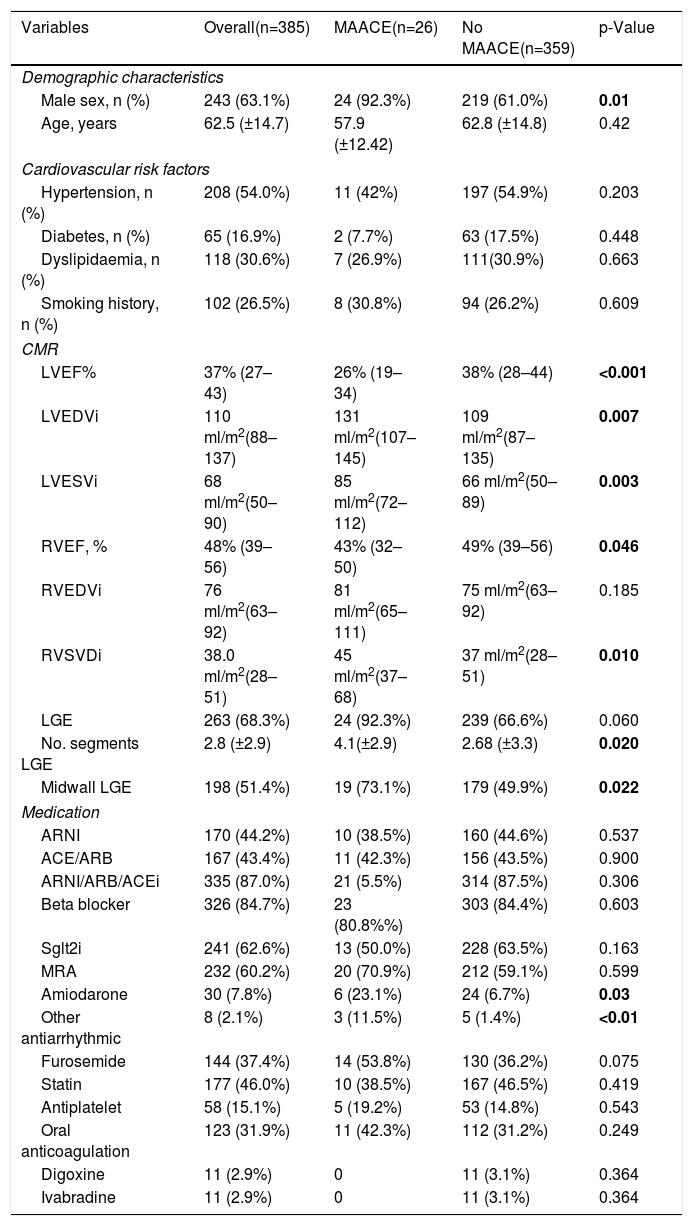
ResultsThe baseline characteristics of the 385 NICM patients are shown in Table 2.
Baseline characteristics and MRI data.
| Variables | Overall(n=385) | MAACE(n=26) | No MAACE(n=359) | p-Value |
|---|---|---|---|---|
| Demographic characteristics | ||||
| Male sex, n (%) | 243 (63.1%) | 24 (92.3%) | 219 (61.0%) | 0.01 |
| Age, years | 62.5 (±14.7) | 57.9 (±12.42) | 62.8 (±14.8) | 0.42 |
| Cardiovascular risk factors | ||||
| Hypertension, n (%) | 208 (54.0%) | 11 (42%) | 197 (54.9%) | 0.203 |
| Diabetes, n (%) | 65 (16.9%) | 2 (7.7%) | 63 (17.5%) | 0.448 |
| Dyslipidaemia, n (%) | 118 (30.6%) | 7 (26.9%) | 111(30.9%) | 0.663 |
| Smoking history, n (%) | 102 (26.5%) | 8 (30.8%) | 94 (26.2%) | 0.609 |
| CMR | ||||
| LVEF% | 37% (27–43) | 26% (19–34) | 38% (28–44) | <0.001 |
| LVEDVi | 110 ml/m2(88–137) | 131 ml/m2(107–145) | 109 ml/m2(87–135) | 0.007 |
| LVESVi | 68 ml/m2(50–90) | 85 ml/m2(72–112) | 66 ml/m2(50–89) | 0.003 |
| RVEF, % | 48% (39–56) | 43% (32–50) | 49% (39–56) | 0.046 |
| RVEDVi | 76 ml/m2(63–92) | 81 ml/m2(65–111) | 75 ml/m2(63–92) | 0.185 |
| RVSVDi | 38.0 ml/m2(28–51) | 45 ml/m2(37–68) | 37 ml/m2(28–51) | 0.010 |
| LGE | 263 (68.3%) | 24 (92.3%) | 239 (66.6%) | 0.060 |
| No. segments LGE | 2.8 (±2.9) | 4.1(±2.9) | 2.68 (±3.3) | 0.020 |
| Midwall LGE | 198 (51.4%) | 19 (73.1%) | 179 (49.9%) | 0.022 |
| Medication | ||||
| ARNI | 170 (44.2%) | 10 (38.5%) | 160 (44.6%) | 0.537 |
| ACE/ARB | 167 (43.4%) | 11 (42.3%) | 156 (43.5%) | 0.900 |
| ARNI/ARB/ACEi | 335 (87.0%) | 21 (5.5%) | 314 (87.5%) | 0.306 |
| Beta blocker | 326 (84.7%) | 23 (80.8%%) | 303 (84.4%) | 0.603 |
| Sglt2i | 241 (62.6%) | 13 (50.0%) | 228 (63.5%) | 0.163 |
| MRA | 232 (60.2%) | 20 (70.9%) | 212 (59.1%) | 0.599 |
| Amiodarone | 30 (7.8%) | 6 (23.1%) | 24 (6.7%) | 0.03 |
| Other antiarrhythmic | 8 (2.1%) | 3 (11.5%) | 5 (1.4%) | <0.01 |
| Furosemide | 144 (37.4%) | 14 (53.8%) | 130 (36.2%) | 0.075 |
| Statin | 177 (46.0%) | 10 (38.5%) | 167 (46.5%) | 0.419 |
| Antiplatelet | 58 (15.1%) | 5 (19.2%) | 53 (14.8%) | 0.543 |
| Oral anticoagulation | 123 (31.9%) | 11 (42.3%) | 112 (31.2%) | 0.249 |
| Digoxine | 11 (2.9%) | 0 | 11 (3.1%) | 0.364 |
| Ivabradine | 11 (2.9%) | 0 | 11 (3.1%) | 0.364 |
Continuous variables presented as mean±standard deviation or median (25th–75th percentiles) where appropriate. ACEi: angiotensin converting enzyme inhibitor; ARB: angiotensin II receptor blocker; ARNI: angiotensin receptor-neprilysin inhibitor; LGE: late gadolinium enhancement; LV: left ventricle; LVEDVi: left ventricular end-diastolic volume index; MRA: mineralocorticoid receptor antagonists; RV: right ventricle; RVEDVi: right ventricular end-diastolic volume index; RVEF: right ventricular ejection fraction; SGLT2i: SGLT2 inhibitor.
Bold values had statistical significance p value <0.05.
A total of 385 patients were included in the study; 63% were male, with a mean age of 62.5 years. The median CMR-derived left ventricular ejection fraction (LVEF) was 37%, and 43% of patients (n=166) had an LVEF <35%. Late gadolinium enhancement (LGE) was present in 68.3% of patients, with a midwall pattern identified in 51.4%.
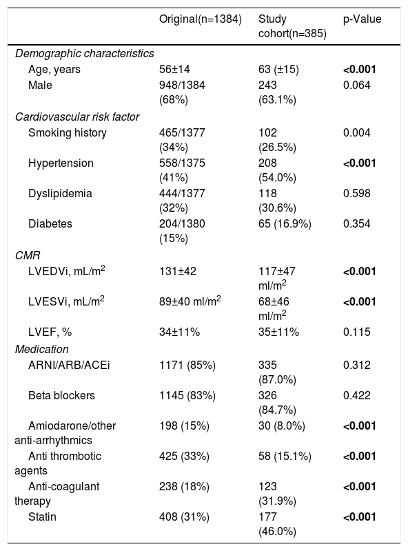
Regarding heart failure pharmacological therapy, 87.0% of patients were treated with renin–angiotensin–aldosterone system (RAAS) inhibitors, and 84.7% received beta-blockers. Use of sodium–glucose cotransporter 2 (SGLT2) inhibitors and mineralocorticoid receptor antagonists (MRAs) was slightly lower, at 62.6% and 60.2%, respectively. Amiodarone was the most frequently prescribed antiarrhythmic drug, used in 7.8% of patients. At the end of follow-up, 102 patients (26.5%) had an implantable cardioverter-defibrillator (ICD) or cardiac resynchronization therapy defibrillator (CRT-D); 5 had a device implanted prior to CMR, and 97 underwent device implantation during follow-up. Eleven patients received cardiac resynchronization therapy pacemakers (CRT-P). When compared with the DERIVATE-NICM cohort (Table 3), the present population was older (63±15 vs. 54±14 years; p<0.001), while the proportion of male patients was similar (63% vs. 68%; p=0.06). This cohort reflects more contemporary heart failure management, with higher use of angiotensin receptor–neprilysin inhibitors (ARNI) and SGLT2 inhibitors, which were not widely prescribed in the original study. However, overall use of RAAS inhibitors and beta-blockers was comparable between cohorts (p=0.312 and p=0.422, respectively). CMR-derived LVEF did not differ significantly between studies (p=0.115), although left ventricular volumes were larger in the DERIVATE-NICM cohort (LVEDVi: 131 mL/m2; p<0.001).
Comparison between baseline characteristic of Derivate NICM study cohort and this Portuguese validation cohort.
| Original(n=1384) | Study cohort(n=385) | p-Value | |
|---|---|---|---|
| Demographic characteristics | |||
| Age, years | 56±14 | 63 (±15) | <0.001 |
| Male | 948/1384 (68%) | 243 (63.1%) | 0.064 |
| Cardiovascular risk factor | |||
| Smoking history | 465/1377 (34%) | 102 (26.5%) | 0.004 |
| Hypertension | 558/1375 (41%) | 208 (54.0%) | <0.001 |
| Dyslipidemia | 444/1377 (32%) | 118 (30.6%) | 0.598 |
| Diabetes | 204/1380 (15%) | 65 (16.9%) | 0.354 |
| CMR | |||
| LVEDVi, mL/m2 | 131±42 | 117±47 ml/m2 | <0.001 |
| LVESVi, mL/m2 | 89±40 ml/m2 | 68±46 ml/m2 | <0.001 |
| LVEF, % | 34±11% | 35±11% | 0.115 |
| Medication | |||
| ARNI/ARB/ACEi | 1171 (85%) | 335 (87.0%) | 0.312 |
| Beta blockers | 1145 (83%) | 326 (84.7%) | 0.422 |
| Amiodarone/other anti-arrhythmics | 198 (15%) | 30 (8.0%) | <0.001 |
| Anti thrombotic agents | 425 (33%) | 58 (15.1%) | <0.001 |
| Anti-coagulant therapy | 238 (18%) | 123 (31.9%) | <0.001 |
| Statin | 408 (31%) | 177 (46.0%) | <0.001 |
ACEi: angiotensin-converting enzyme inhibitor; ARB: angiotensin receptor blocker; ARNI: angiotensin receptor-neprilysin inhibitor.
Bold values had statistical significance p value <0.05.
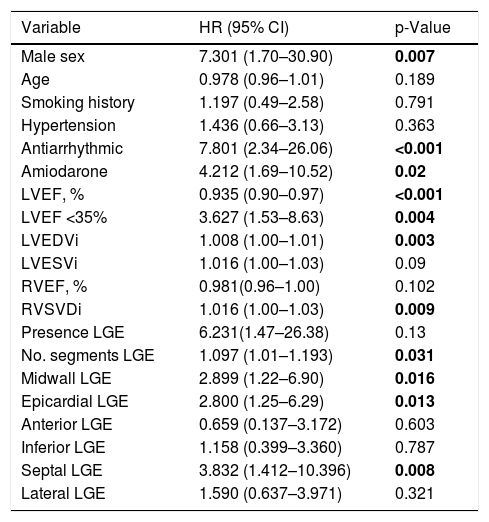
Univariable analysis (Table 4) identified male sex and the use of amiodarone or other antiarrhythmic drugs as clinical variables statistically associated with major arrhythmic cardiac events (MAACE). Among CMR-derived parameters, lower left ventricular ejection fraction (LVEF) and higher left ventricular end-diastolic volume indexed (LVEDVi) were also associated with arrhythmic events. With respect to tissue characterization, the presence of late gadolinium enhancement (LGE) per se was not associated with MAACE; however, both the extent of LGE, expressed as the number of involved segments, specific LGE patterns – namely midwall or epicardial, and septal involvement were significantly associated with arrhythmic outcomes.
Univariable predictors of MAACE.
| Variable | HR (95% CI) | p-Value |
|---|---|---|
| Male sex | 7.301 (1.70–30.90) | 0.007 |
| Age | 0.978 (0.96–1.01) | 0.189 |
| Smoking history | 1.197 (0.49–2.58) | 0.791 |
| Hypertension | 1.436 (0.66–3.13) | 0.363 |
| Antiarrhythmic | 7.801 (2.34–26.06) | <0.001 |
| Amiodarone | 4.212 (1.69–10.52) | 0.02 |
| LVEF, % | 0.935 (0.90–0.97) | <0.001 |
| LVEF <35% | 3.627 (1.53–8.63) | 0.004 |
| LVEDVi | 1.008 (1.00–1.01) | 0.003 |
| LVESVi | 1.016 (1.00–1.03) | 0.09 |
| RVEF, % | 0.981(0.96–1.00) | 0.102 |
| RVSVDi | 1.016 (1.00–1.03) | 0.009 |
| Presence LGE | 6.231(1.47–26.38) | 0.13 |
| No. segments LGE | 1.097 (1.01–1.193) | 0.031 |
| Midwall LGE | 2.899 (1.22–6.90) | 0.016 |
| Epicardial LGE | 2.800 (1.25–6.29) | 0.013 |
| Anterior LGE | 0.659 (0.137–3.172) | 0.603 |
| Inferior LGE | 1.158 (0.399–3.360) | 0.787 |
| Septal LGE | 3.832 (1.412–10.396) | 0.008 |
| Lateral LGE | 1.590 (0.637–3.971) | 0.321 |
LGE: late gadolinium enhancement; LVEF: left ventricular ejection fraction.
Bold values had statistical significance p value <0.05.
The median DERIVATE Risk Score 2.0 (DRS 2.0) was 6.0 (interquartile range: 4.0–9.0). According to predefined risk categories, 20% of patients were classified as low risk (n=76), 53% as intermediate risk (n=203), and 28% as high risk (n=106).
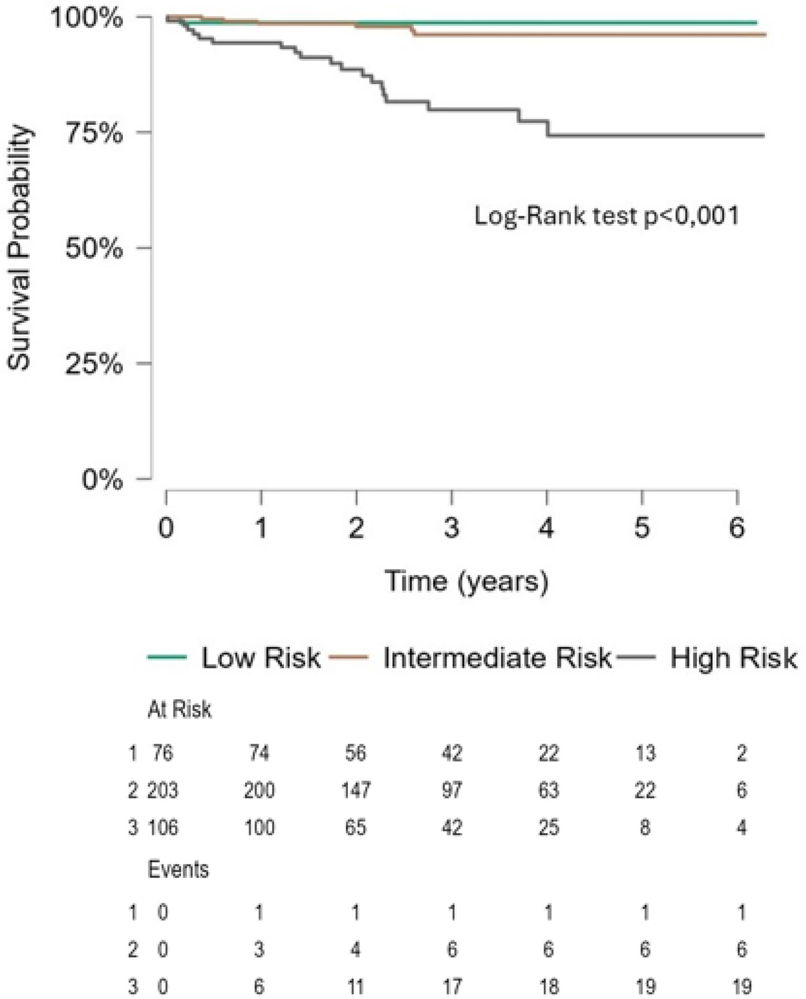
Over a mean follow-up of 3.0±1.4 years, 26 MAACE (6.8%) were observed, including seven sudden cardiac deaths, nine episodes of sustained ventricular tachycardia, and 10 appropriate implantable cardioverter-defibrillator therapies. Annualized MAACE rates increased progressively across risk categories, at 0.4%/year in the low-risk group, 1.0%/year in the intermediate-risk group, and 6.0%/year in the high-risk group. Patients classified as high risk by the DRS 2.0 had an eightfold higher risk of MAACE compared with those in the low- or intermediate-risk categories (hazard ratio 8.0; 95% confidence interval 3.36–19.04; p<0.001).
Event-free survival according to DRS 2.0 risk categories is illustrated in the Kaplan–Meier curves (Figure 1). At 3 years of follow-up, estimated event-free survival was 98.8%, 97.1%, and 82.1% in the low-, intermediate-, and high-risk groups, respectively.

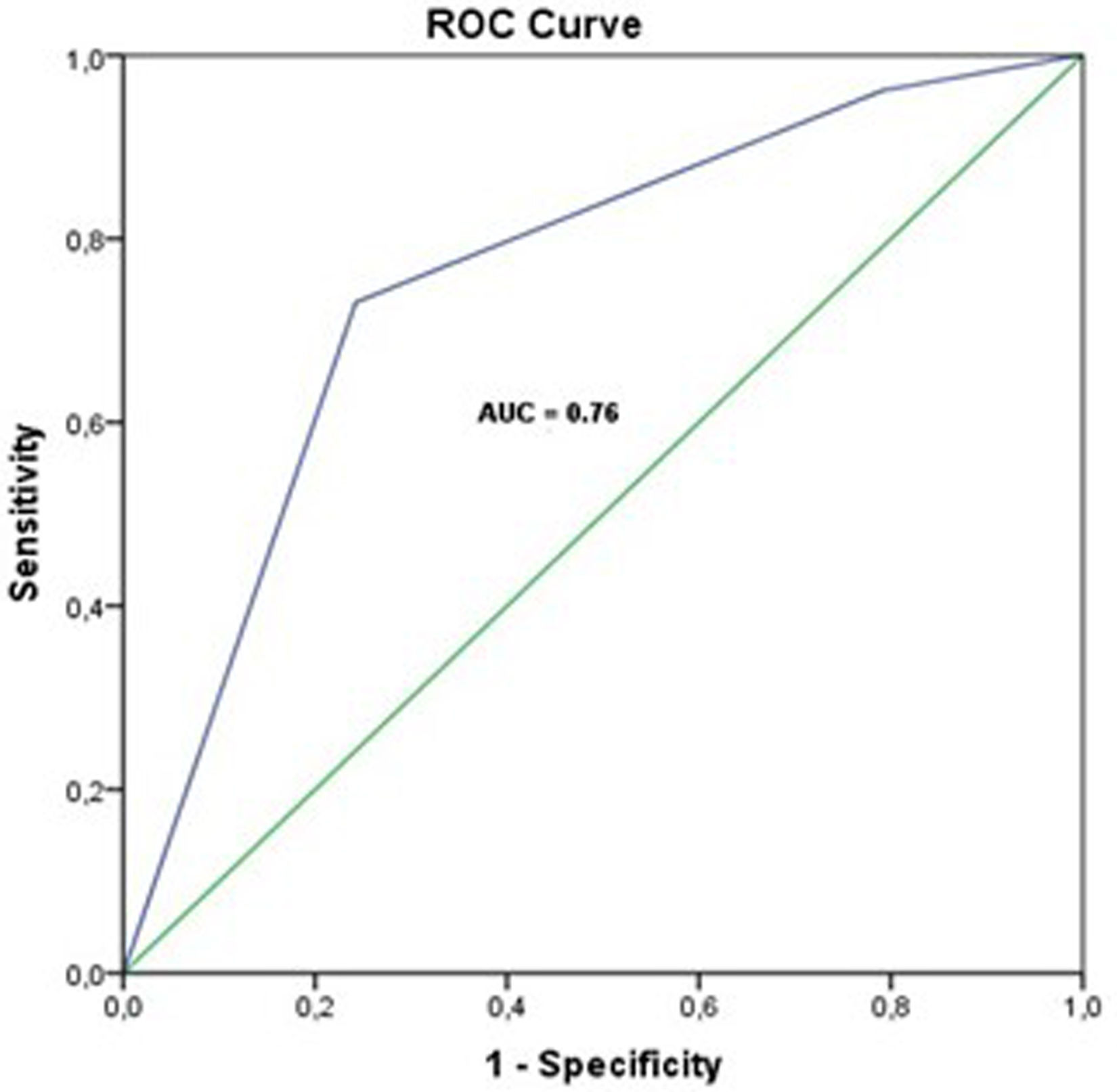
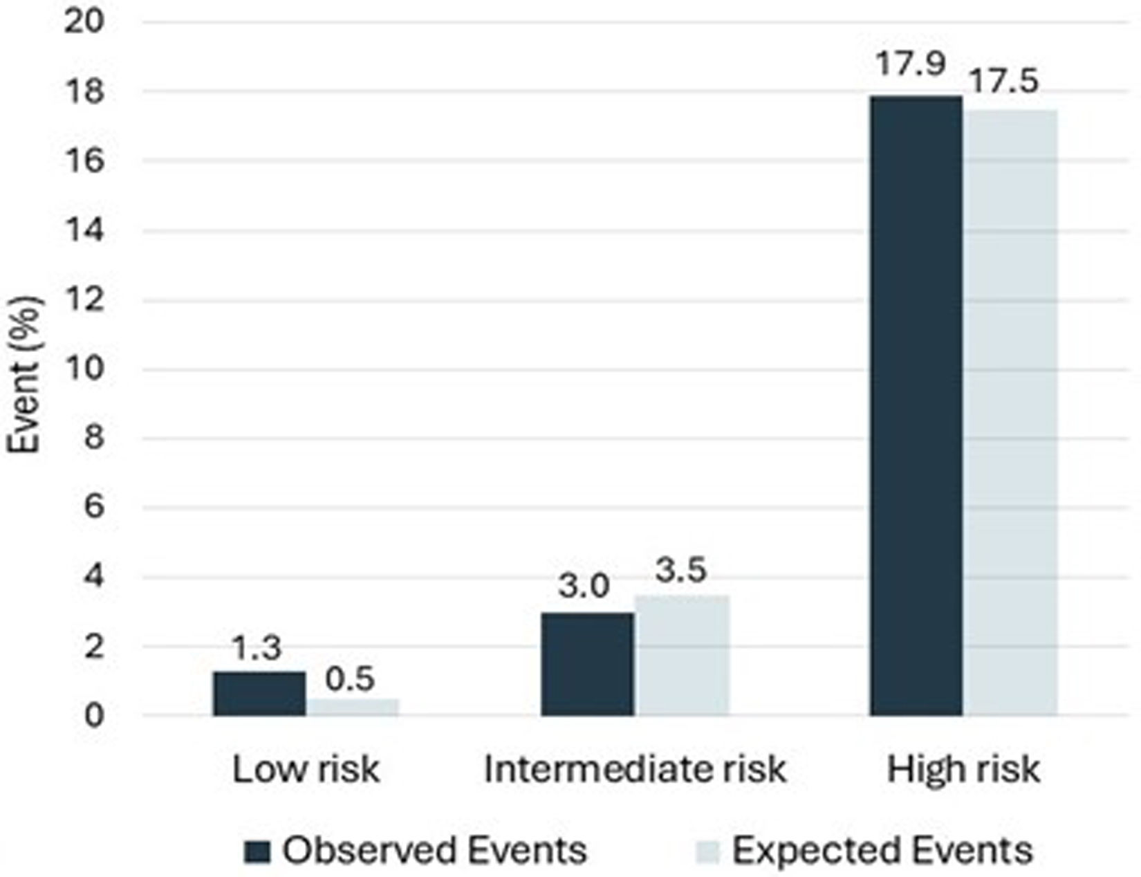
The DRS 2.0 demonstrated good discriminative ability, with a C-statistic of 0.76 (95% confidence interval 0.66–0.85; p<0.001) (Figure 2). Model calibration was satisfactory, as indicated by a non-significant Hosmer–Lemeshow goodness-of-fit test (χ2=0.802; p=0.371) (Figure 3).


In this contemporary cohort of patients in Portugal with non-ischemic cardiomyopathy (NICM), application of the DERIVATE Risk Score 2.0 (DRS 2.0) enabled stratification of arrhythmic risk across clinically distinct groups. A minority of patients classified as high risk accounted for most major arrhythmic cardiac events (MAACE), whereas most patients in the low- and intermediate-risk categories remained event-free during follow-up. These findings suggest that the score retains meaningful discriminatory capacity in this setting and may provide incremental clinical insight beyond conventional risk markers.
The overall incidence of arrhythmic events in this cohort was low, with an annual MAACE rate of 2.3% and a sudden cardiac death (SCD) rate of 0.6% per year. These results are consistent with contemporary data and likely reflect substantial advances in heart failure management. In the DANISH trial, for example, the annual SCD rate was 1.13% despite inclusion of patients with left ventricular ejection fraction (LVEF) <35%.2 The low event burden observed in this study is plausibly explained by high uptake of guideline-directed medical therapy (GDMT), as well as the use of cardiac resynchronization therapy in a subset of patients.
More than 80% of patients in this cohort were treated with renin–angiotensin system inhibitors and beta-blockers, while approximately 60% received mineralocorticoid receptor antagonists and sodium–glucose cotransporter 2 inhibitors. Although these figures remain slightly below those reported in randomized clinical trials such as DAPA-HF and EMPEROR-Reduced,11,12 they compare favorably with other real-world observational studies, underscoring the contemporary nature of this cohort.13,14 Notably, while 43% of patients had a CMR-derived LVEF <35%, only 26% ultimately received an implantable cardioverter-defibrillator (ICD) or CRT-D, closely mirroring the proportion of patients classified as high risk by the DRS 2.0. This discrepancy may reflect recovery of LVEF in some patients during follow-up, but it may also indicate that clinical decision-making is increasingly influenced by factors beyond LVEF alone.
The DRS 2.0 integrates simple clinical variables with advanced CMR-derived measures of ventricular function and myocardial tissue characterization. By incorporating not only the presence of late gadolinium enhancement (LGE), but also its extent and regional distribution, the score captures key features of the arrhythmogenic substrate in NICM more comprehensively than binary LGE assessment. This approach aligns with growing evidence that both the burden and anatomical pattern of myocardial fibrosis are critical determinants of arrhythmic risk.15,16
In recent years, several alternative models have been proposed to refine arrhythmic risk stratification in NICM, many of which include increasingly sophisticated LGE-based metrics, such as detailed quantification, three-dimensional remodeling indices, or measures of fibrosis heterogeneity.17,18 While these approaches are promising, their clinical applicability may be limited by technical complexity, lack of standardization, or restricted availability outside specialized centers. In this context, risk scores that balance prognostic performance with clinical feasibility remain particularly relevant.19
Importantly, validation of clinical risk scores in populations distinct from those in which they were derived is essential before widespread clinical adoption. Differences in genetic background, disease etiology, comorbidity burden, healthcare systems, referral patterns, and therapeutic strategies can all influence baseline risk and modify the performance of prognostic models. Scores developed in selected trial populations or highly specialized centers may demonstrate attenuated discrimination or miscalibration when applied to broader or different real-world cohorts.20 External validation therefore serves not only to confirm generalizability, but also to identify potential limitations, recalibration needs, or subgroups in which a given model may perform suboptimally.21
Several limitations of the present study should be acknowledged. Its single-center observational design and the relatively small number of arrhythmic events limited statistical power and precluded multivariable analysis. In addition, although these findings support the applicability of the DRS 2.0 in a Southern European cohort, further prospective validation in larger and more diverse populations is required. Future developments in risk stratification may also benefit from the integration of genotype-specific data, particularly for variants known to confer elevated arrhythmic risk in NICM.22,23
ConclusionIn this contemporary real-world cohort of patients with non-ischemic cardiomyopathy, the DERIVATE Risk Score 2.0 demonstrated the ability to stratify arrhythmic risk across clinically distinct groups. Despite low overall event rates in the setting of optimized guideline-directed medical therapy, the score identified a subset of patients with a disproportionate burden of major arrhythmic events. While further multicenter validation and incorporation of genetic data are warranted, these findings suggest that multiparametric risk models such as the DRS 2.0 may complement conventional LVEF-based approaches in refining arrhythmic risk assessment.
Ethical statementThis study was performed in line with the principles of the Declaration of Helsinki. The requirement for informed consent was waived due to the retrospective nature of the study and the use of de-identified patient data. All data were anonymized prior to analysis to ensure patient confidentiality. Access to the electronic medical records was restricted to the research team. The authors declare that they have no known competing financial interests.
Conflicts of interestThe authors have no conflicts of interest to declare.