Patients with Fontan circulation may present desaturation on effort. The main causes are previously created fenestrations, pulmonary arteriovenous malformations, venovenous collaterals, and coronary sinus to atrium communications.1–3 An extremely rare but important cause is Fontan to atrium communication, which can be treated percutaneously.4
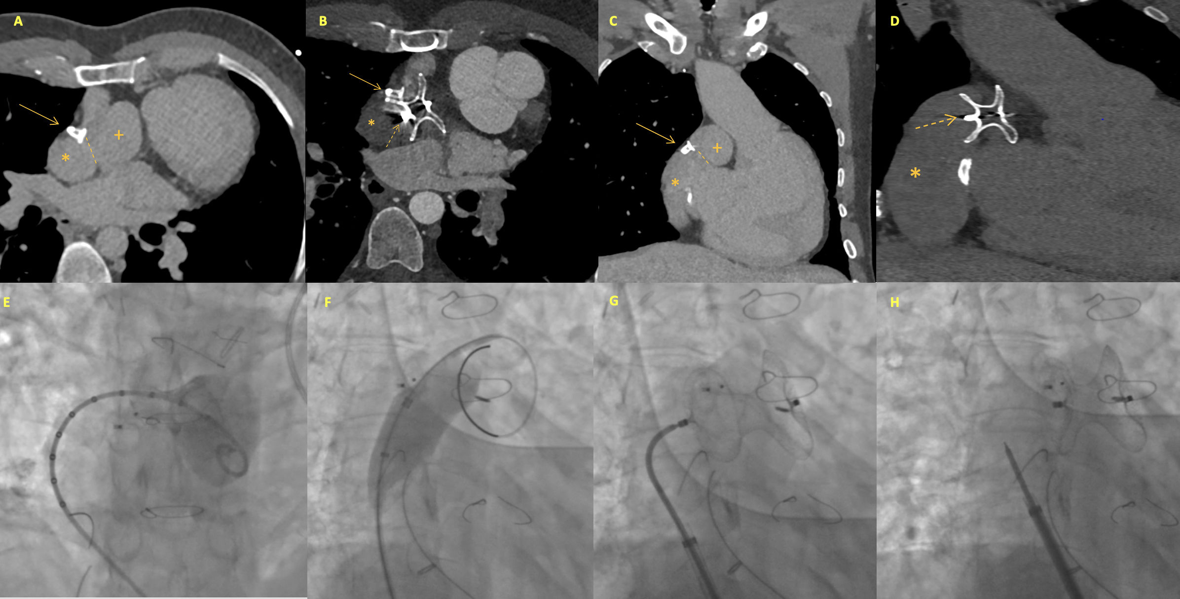
We present the case of a 33-year-old man with an unbalanced atrioventricular canal treated with fenestrated Fontan palliation, which subsequently required percutaneous fenestration closure and atrial septal defect (ASD) closure with an Amplatzer device. He underwent right heart catheterization due to baseline desaturation worsened by exercise. Although mean pressure in the Fontan system was normal (10 mmHg), a right-to-left shunt was detected between the Fontan and a pseudocavity connected to the systemic atrium. A preprocedural computed tomography (CT) angiogram performed in venous and arterial phases showed communication between the Fontan conduit (*) and a 2.7 cm×2.6 cm pseudocavity (+) located adjacent to the previously placed ASD closure device (Figure 1A and C), suggestive of an atrial pseudoaneurysm. Transcatheter closure was attempted, and an angiogram confirmed the connection (Figure 1E). The neck of the connection was sized using a 25 mm balloon. Given the presence of an adequate rim for anchoring, and in order to achieve complete sealing of the orifice, a 24 mm Amplatzer atrial septal occluder was used to close the shunt (Figure 1F and G). No pressure increase was noted in the Fontan circuit before delivering the device (Figure 1H), and baseline saturation improved from 88% to 95%. The patient remained asymptomatic six months post-procedure, with normal baseline and exercise saturation. A follow-up CT showed that the ASD occluder was well-positioned (discontinuous arrow), with complete occlusion of the cavity in both venous and arterial phases (Figure 1B and D).

Preprocedural imaging (A and C): communication between the Fontan conduit (*) and a pseudocavity (+) located adjacent to the previously placed ASD closure device (continuous arrow), with an entry point (dashed lines) measuring 1.2 cm in maximum diameter. Intraprocedural angiography and intervention (E–H): after confirming the presence of the connection, balloon sizing was performed using a 25 mm balloon (waist diameter 16 mm) and a 24 mm Amplatzer ASD occluder was deployed. No increase in Fontan pressures was confirmed prior to device release. Follow-up CT (B and D): a well-positioned ASD occluder (discontinuous arrow), juxtaposed to the previous device (continuous arrow), with complete occlusion of the cavity.
None declared.
Conflicts of interestNone declared.
Data availabilityThe data underlying this article will be shared on reasonable request to the corresponding author.