
Sugestões
Idioma
Informação da revista
Vol. 45. Núm. 10.
Páginas 553-610 (Outubro 2026)
Citação
Citação
Partilhar
Baixar PDF
Mais opções do artigo
Vol. 45. Núm. 10.
Páginas 553-610 (Outubro 2026)
Image in Cardiology
Acesso de texto completo

Massive pneumopericardium caused by perforated gastric ulcer

Pneumopericárdio maciço causado por úlcera gástrica perfurada
Visitas
734
Roberto Ramos Barbosa
Autor para correspondência
roberto.cardio@gmail.com

Corresponding author.
, Fernando Henrique Rabelo Abreu dos Santos, Guilherme Carvalho Moitinho, Marcio Rufino Barbosa Junior, Karolini Zambelli Marques Alves, Pamella Souza Macedo, Luiz Fernando Machado Barbosa
Hospital da Santa Casa de Misericórdia de Vitória, Vitória, ES, Brazil
Este item recebeu
Informação do artigo
Texto Completo
Bibliografia
Baixar PDF
Estatísticas
Figuras (1)
fig0005
Texto Completo

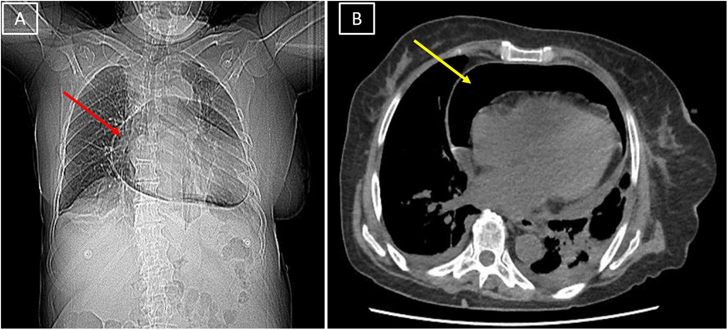
A 66-year-old woman was admitted to the emergency department presenting with dyspnea and bilateral chest pain radiating to the back and normal blood pressure, but with impaired peripheral perfusion and sinus tachycardia. Initial electrocardiogram showed right bundle branch block and no signs of myocardial ischemia. Chest X-ray followed by a computed tomography scan revealed a large pneumopericardium (Figure 1). After initial stabilization, diagnostic upper digestive endoscopy was urgently performed, which revealed a gastric ulcer suggestive of malignancy, with perforation directed towards the pericardium.

Figure 1.

(A) Chest X-ray showing enlargement of the cardiac area and pericardial thickening with the presence of gaseous content (red arrow). (B) Chest computed tomography scan showing a large amount of air in the pericardial sac, confirming the diagnosis of pneumopericardium (yellow arrow).

Firstly, bed side pericardiocentesis was performed. Then, a multidisciplinary team decided upon surgical intervention. Intraoperative view identified a paraoesophageal hernia with hiatal widening and a perforated ulcer firmly adhered to the thickened pericardium. Partial gastrectomy of the perforated segment was performed, with proximal gastric tubulization. Due to esophageal retraction, part of the tubulized stomach was located in the mediastinum. Thoracic and double mediastinal drainage were performed, and a post-pyloric nasoenteric tube was inserted. Despite successful surgical outcome, the patient was readmitted for pneumonia and died 68 days later. Histopathological analysis revealed an ischemic type Cameron ulcer.

Chest pain is a recurrent symptom in the emergency department, and unstable clinical presentations must alert us to acute critical conditions.1,2 A perforated gastric ulcer is a severe and life-threatening medical emergency.3,4 Spontaneous perforations with systemic changes are uncommon.4 This rare, massive and typical pneumopericardium5 seen on chest imaging initiated an extensive clinical reasoning and individualized decision making. Unfortunately, respiratory complications led to death in the short term after a challenging operation.

Ethical approval

Santa Casa de Misericórdia de Vitória Higher School of Sciences – EMESCAM. Approval number: 6,091,402.

Conflicts of interest

None declared.

References
[1]
M. Gulati, P.D. Levy, D. Mukherjee, et al.
2021 AHA/ACC/ASE/CHEST/SAEM/SCCT/SCMR guideline for the evaluation and diagnosis of chest pain: a report of the American College of Cardiology/American Heart Association Joint Committee on Clinical Practice Guidelines.
Circulation, 144 (2021), pp. e368-e454
[2]
J. Ouachaou, I. Laaribi, H. Mimouni, et al.
Post-traumatic compressive pneumopericardium with spontaneous ventilation: case report.
Respir Med Case Rep, 32 (2021),
[3]
K. Søreide, K. Thorsen, E.M. Harrison, et al.
Perforated peptic ulcer.
Lancet, 386 (2015), pp. 1288-1298
[4]
R. Tanaka, S. Kosugi, K. Sakamoto, et al.
Treatment for perforated gastric ulcer: a multi-institutional retrospective review.
J Gastrointest Surg, 17 (2013), pp. 2074-2081
[5]
A.S. Osuna Garibay, C. Arroyo-Rodríguez, J.R. Victoria-Nandayapa, et al.
Tension pneumopericardium secondary to esophageal-pericardial fistula.
Rev Port Cardiol, 41 (2022), pp. 977-978
Copyright © 2026. Sociedade Portuguesa de Cardiologia
Baixar PDF
Idiomas
Revista Portuguesa de Cardiologia
Opções de artigo
Ferramentas