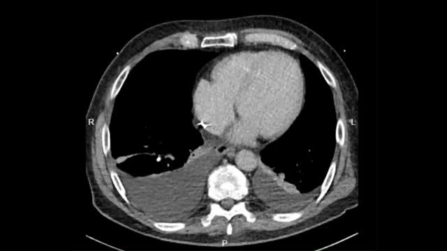
A 72-year-old man presented with inflammatory signs and partial exteriorization of a dual-chamber pacemaker (PM) generator implanted 15 years previously for Mobitz II atrioventricular block. One year after the implantation, a new ventricular lead was implanted due to dysfunction, leaving the old one abandoned; the generator was replaced 9 years later. The device was programmed in DDDR pacing.
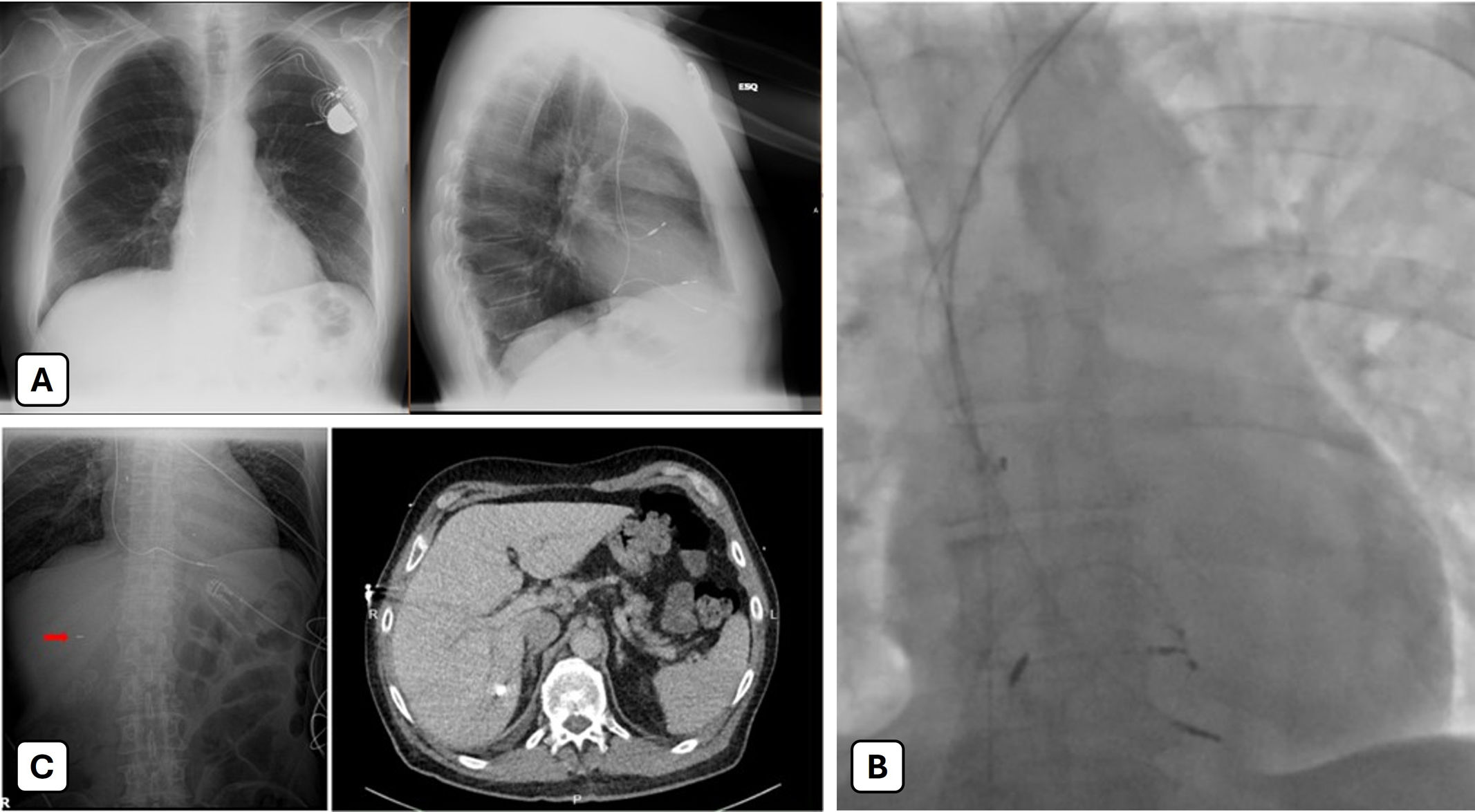
On admission, laboratory tests showed leukocytosis, neutrophilia, and elevated C-reactive protein. A chest X-ray confirmed the presence of one atrial and two ventricular leads (Figure 1A). Transesophageal echocardiography revealed leads in situ, no vegetations, and no pericardial effusion. PM interrogation documented episodes of paroxysmal atrial fibrillation with >90% ventricular pacing. Cultures from the purulent pocket grew methicillin-sensitive Staphylococcus aureus and Corynebacterium, while blood cultures remained negative. At this point, a diagnosis of pocket infection without bacteremia was made. After 15 days of targeted antibiotic therapy, the patient was referred for extraction.

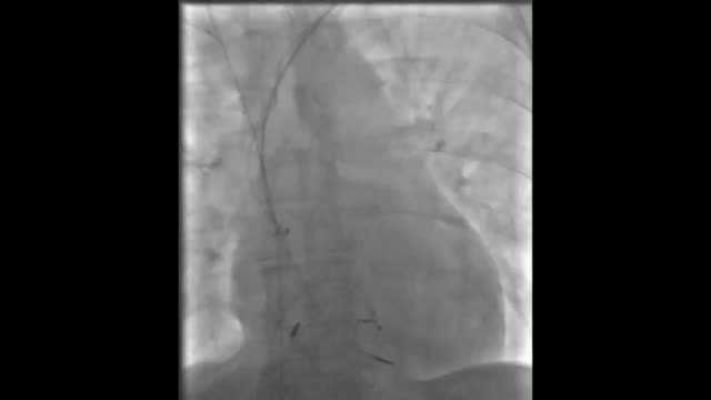
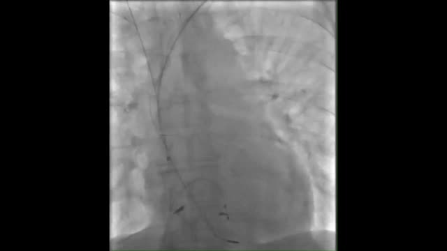
Following extensive debridement at the generator site, the proximal portions of both ventricular leads and the right atrial lead were isolated. Extraction was initiated through the atrial lead (an old passive lead), using polypropylene telescoping non-powered countertraction sheaths (7, 8.5, and 10F) with the PISA technique. This lead was gradually freed from fibrotic adhesions until it fractured, leaving the tip attached to the myocardium. The metallic ring detached from the tip and migrated into the right atrium (Figure 1B and Supplemental Video 1). During subsequent removal of the abandoned ventricular lead, the metallic ring migrated further into a suprahepatic vein, where it remained (Supplemental Video 2). Both ventricular leads were ultimately and completely extracted with the PISA technique, and a temporary active-fixation lead was implanted as a bridge to reimplantation.
The patient remained asymptomatic. Postprocedural imaging demonstrated the metallic ring lodged in hepatic segment VII (Figure 1C and Supplemental Video 3); there was no hepatic dysfunction or cytolysis. Cultures confirmed S. aureus in one extracted RV lead and pocket samples, whereas the abandoned lead and the atrial lead were sterile. A multidisciplinary team, including interventional radiology and hepatology, reviewed the case. Given the patient's stability, preserved liver function, and lack of ongoing infection, removal of the migrated fragment was deemed unnecessary, and conservative management was chosen. Antibiotics were continued until blood cultures cleared.
Lead fragment embolization is a rare complication of transvenous extraction, occurring in approximately 0.2% of procedures, most often in the pulmonary vasculature.1–4 Asymptomatic fragments are generally managed conservatively, with retrieval reserved for patients who are symptomatic or develop organ dysfunction.4
To our knowledge, this is the first reported case of embolization of a lead fragment to the liver. Careful evaluation of risks, benefits, and clinical context is essential when considering the retrieval of migrated fragments.
FundingThere was no funding for this article.
Conflicts of interestThe authors have no conflicts of interest to declare.
The following are the supplementary data to this article: