

Lipoprotein(a) [Lp(a)] is recognized as an independent risk factor for coronary disease owing to its atherogenic, proinflammatory, and prothrombotic properties. Current guidelines recommend a single measurement in adults to refine cardiovascular (CV) risk assessment. We aimed to characterize Lp(a) levels in patients with acute coronary syndrome (ACS) and explore associations with sex, age, comorbidities, and traditional cardiovascular risk factors.
MethodsWe conducted a cross-sectional study of patients with ACS admitted to our center between January 2022 and December 2023, with Lp(a) measured at admission. Patients were stratified into two groups: Lp(a) >100 nmol/L and ≤100 nmol/L. Demographic and clinical data, including traditional cardiovascular risk factors (dyslipidemia, diabetes mellitus, arterial hypertension, smoking, and obesity), were collected from hospital records. Chi-square and independent T or Mann–Whitney U tests were used to compare categorical and quantitative variables; linear regression analysis assessed associations between continuous Lp(a) values and independent variables.
ResultsAmong 903 patients admitted with ACS during the study period, Lp(a) was measured in 388 (42%). Median Lp(a) level was 62.0 [18.4, 153.8] nmol/L. Of these, 38.7% had Lp(a) >100 nmol/L. Women had higher Lp(a) than men (p-trend=0.014). Lp(a) levels were similar across traditional cardiovascular risk factors categories. Among patients without traditional cardiovascular risk factors, women also had higher Lp(a) than men (p=0.003). Elevated Lp(a) was associated with a history of coronary artery disease (p-trend=0.003) and with treatment with high-intensity statins alone (p-trend=0.032) or in combination with ezetimibe (p-trend=0.014).
ConclusionsLp(a) levels showed a heterogeneous distribution and were not associated with traditional cardiovascular risk factors or other lipid parameters. This reinforces Lp(a) as an independent risk factor, supporting active screening in patients with ACS, particularly in women not affected by traditional cardiovascular risk factors.
A lipoproteína(a) [Lp(a)] é um fator de risco independente para doença coronária, pelas suas propriedades aterogénicas, pró-inflamatórias e protrombóticas. As diretrizes atuais recomendam uma medição única em adultos para melhor estratificação do risco cardiovascular. Este estudo visa caracterizar os níveis de Lp(a) em doentes com síndrome coronária aguda (SCA) e analisar associações com sexo, idade, comorbilidades e fatores de risco cardiovascular tradicionais (FRCVT).
MétodosForam incluídos doentes com SCA admitidos no nosso centro entre janeiro de 2022 e dezembro de 2023, com medição da Lp(a) à admissão. Os doentes foram estratificados em dois grupos: Lp(a)>100nmol/L e ≤100nmol/L. Os dados clínicos e demográficos foram extraídos dos registos hospitalares incluindo os FRCVT. As variáveis categóricas e quantitativas foram comparadas com os testes de Qui-quadrado, t de Student ou Mann-Whitney. A regressão linear foi utilizada para avaliar associações entre os valores contínuos de Lp(a) e as variáveis independentes.
ResultadosEntre os 903 doentes internados por SCA durante o período do estudo, a Lp(a) foi medida em 388 (42%). A mediana de Lp(a) foi de 62,0 [18,4, 153,8] nmol/L e 38,7% tinham Lp(a)>100nmol/L. A mulheres apresentaram níveis de Lp(a) superiores aos dos homens (p-trend=0,014). Os níveis de Lp(a) foram semelhantes entre as categorias FRCVT, mas dos doentes sem FRCVT, as mulheres apresentaram Lp(a) superior à dos homens (p=0,003). Níveis elevados de Lp(a) foram associados a doença arterial coronária prévia (p-trend=0,003) e ao uso de estatinas de alta intensidade isoladamente (p-trend=0,032) ou em combinação com ezetimiba (p-trend=0,014).
ConclusõesA Lp(a) apresentou uma distribuição heterogénea, mas com tendência para níveis mais elevados em mulheres com SCA. Estes achados reforçam a necessidade de rastreio da Lp(a) independente dos FRCVT ou comorbilidades, sobretudo em doentes do sexo feminino.

Over the years, strategies for reducing cardiovascular (CV) risk have been recommended and implemented in patients with history of cardiovascular disease (CVD), leading to a global decline in mortality associated with coronary artery disease (CAD).1 However, despite the development and implementation of secondary prevention programs, the global burden of CAD continues to rise, along with its associated morbidity.2 In response to this, European and American Cardiology societies recommend increasingly stringent targets for controlling cardiovascular risk factors following an ischemic event.3–5 Furthermore, in recent years, significant efforts have been made to identify new biomarkers which could improve risk stratification and reduce the burden of CVD.6
Dyslipidemia remains a major risk factor in the pathogenic pathway of patients with CAD.3,7 The EUROASPIRE V Survey, which enrolled 7824 patients from 27 countries, reported that 71% of patients with established CAD had low-density lipoprotein cholesterol (LDL-C) levels ≥1.8 mmol/L (70 mg/dL) despite being on lipid-lowering therapies, thus failing to achieve the recommended targets.7 In this context, other lipid particles beyond LDL-C were investigated, including lipoprotein(a) [Lp(a)], which emerged as a key contributor to residual CV risk.8 Lp(a) is a plasma lipoprotein consisting of a cholesterol-rich LDL particle with one molecule of apolipoprotein B100 and a molecule of apolipoprotein(a) [Apo(a)], linked via a disulfide bond.9 Previous studies have shown that Lp(a) levels are genetically determined in more than 90% of cases.8,10,11 The LPA gene is thought to be fully expressed by the age of two, with adult Lp(a) levels typically established by the age of five, although levels may continue to rise throughout adulthood. Consequently, the European Atherosclerosis Society (EAS) recommends at least one Lp(a) measurement in every adult for CV risk stratification.10
More recently, Lp(a) has been recognized as an independent CVRF for atherosclerotic cardiovascular disease (ASCVD) due to its atherogenic, proinflammatory, and prothrombotic properties, which may even exceed those of LDL-C.9,12 In fact, elevated Lp(a) levels can be a causal risk factor for CVD in approximately 20% of the global population and Lp(a)-lowering therapies are currently under investigation, positioning this plasmatic lipoprotein as a novel target for ASCVD treatment.8,13
Although its pathogenic role is increasingly acknowledged, the extent to which Lp(a) levels influence CV outcomes – particularly in the setting of acute coronary syndrome (ACS) – remains under investigation. Recent evidence has demonstrated that elevated Lp(a) levels are independently associated with long-term major adverse cardiovascular events (MACE) in individuals with and without established ASCVD. Additionally, optimal Lp(a) threshold for risk assessment differs between primary and secondary prevention populations, while emerging evidence highlights Lp(a)’s critical role in triggering and exacerbating acute ischemic events, particularly ACS.14
ObjectivesThis study employed a cross-sectional analysis to characterize Lp(a) levels across a diverse cohort of ACS patients and explore associations with sex, age, comorbidities, and traditional cardiovascular risk factors.
MethodsStudy designWe conducted a cross-sectional study that included all patients admitted with ACS to the Cardiology Department of a tertiary referral hospital (Unidade Local de Saúde Gaia/Espinho) from January 2022 to December 2023. Lp(a) levels were assessed upon admission, and patients were divided into two groups: those with elevated Lp(a) (>100 nmol/L) and those with lower Lp(a) (≤100 nmol/L) levels. Beyond this dichotomous classification, we examined the continuous relationship between Lp(a) concentration and outcomes. For clinical clarity, we set a reference threshold of 100 nmol/L – a value increasingly acknowledged in cardiology literature as clinically significant – allowing us to compare patients with elevated levels to those with concentrations at or below this marker.11
Lp(a) measurement and outcomesSince January 2022, Lp(a) measurements have been opportunistically included in the standard analytical workup for all ACS patients admitted to our hospital. The decision to request Lp(a) testing was made by the attending cardiologists during the initial assessment upon hospital admission. This assessment was performed alongside routine blood tests, including complete blood count, renal function, liver enzymes, lipid profile, glycated hemoglobin (HbA1c), and thyroid function.
Lp(a) levels were measured at our center's laboratory using a particle-enhanced turbidimetric immunoassay, conducted on the Roche Cobas® Integra Z701 analyzer, in accordance with the manufacturer's instructions. The results are expressed in nanomoles per litre (nmol/L).
For the primary analysis, an Lp(a) cut-off of 100 nmol/L was used to define elevated Lp(a) levels, in accordance with the recommendations of the National Lipid Association (NLA) guidelines.11 Secondary analyses explored additional thresholds of 75 nmol/L and 175 nmol/L to assess the sensitivity of findings across clinically relevant cut-off points: 75 nmol/L representing a more conservative threshold suggested for primary prevention populations, and 175 nmol/L corresponding to the eligibility criterion for emerging Lp(a)-lowering therapies in clinical trials.15
VariablesPatient demographic and clinical data were extracted from hospital records. Diabetes mellitus, arterial hypertension, obesity, smoking, and dyslipidemia (defined as LDL-cholesterol levels ≥130 mg/dL) were considered traditional cardiovascular risk factors. Premature CAD was defined as occurring in men aged ≤55 years and women aged ≤60 years.16
Statistical analysisCategorical variables were described as counts and percentages. Continuous variables were described as median and interquartile range (IQR). Valid percentages were reported, and missing data were not imputed.
Lp(a) group comparisons were made using the Chi-squared test for categorical variables and Independent T or Mann–Whitney U tests for continuous variables.
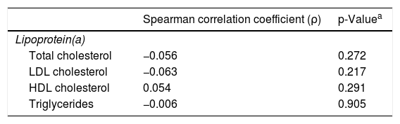
Spearman's correlation coefficients were used to evaluate the relationships between Lp(a) levels and other lipid parameters.
We used R software (version 4.1.2) for all statistical analyses; this meant the data were analyzed using both numerical and graphical methods. Additionally, a p-trend was computed to evaluate the associations between continuous Lp(a) values and independent variables using linear regression analysis. This approach allowed us to assess the direction and significance of potential dose-response relationships rather than simply comparing categorical groups, providing insight into whether higher Lp(a) levels were associated with linear changes in the clinical variables of interest. Statistical significance was set at p<0.05.
Ethical approvalThis study was approved by the Ethics Committee of Unidade Local de Saúde Gaia e Espinho (document number 134/2024).
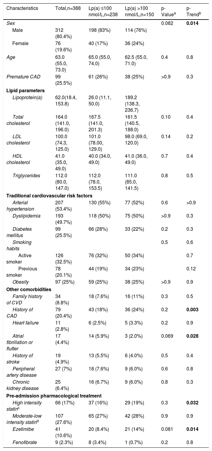
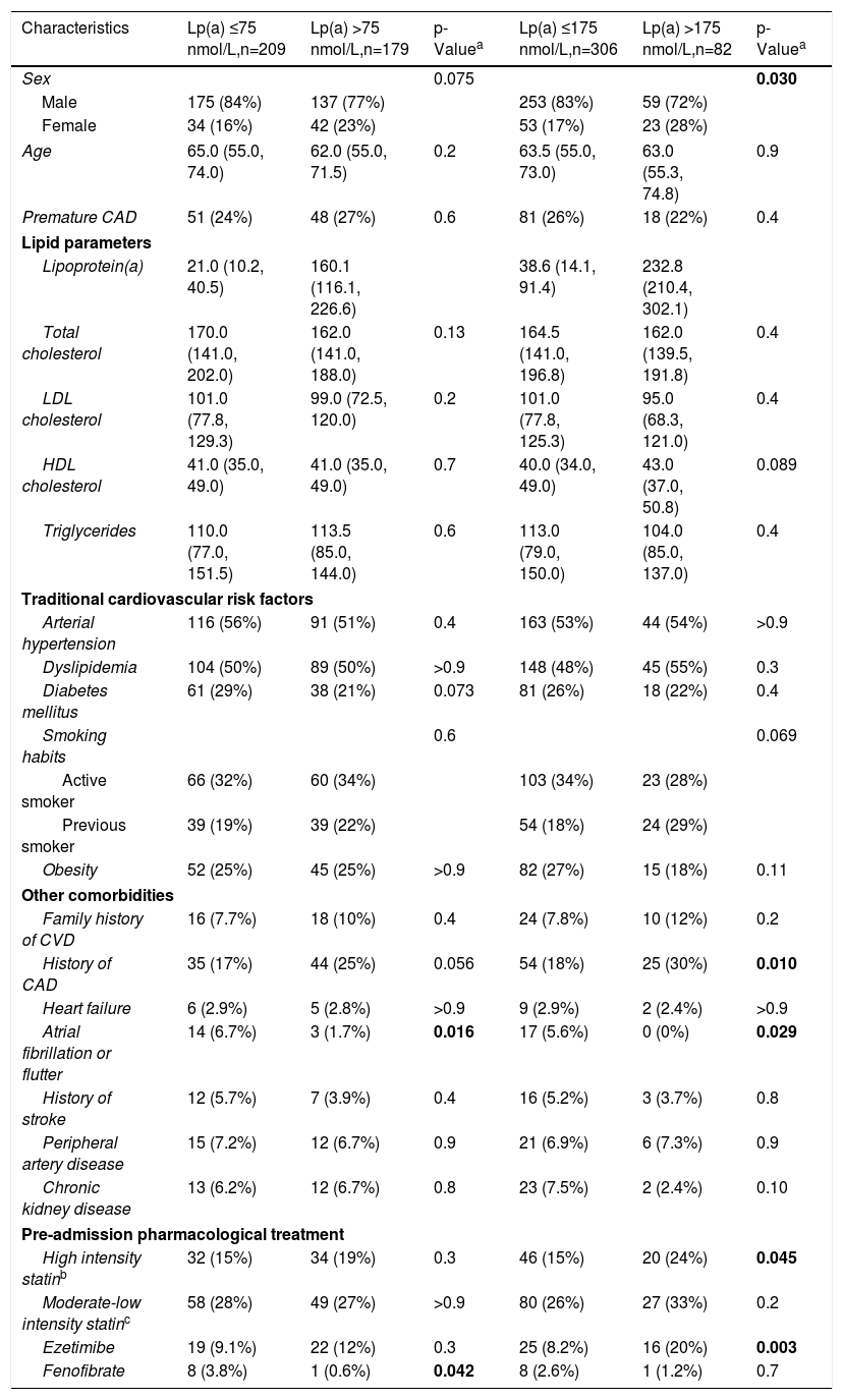
ResultsA total of 903 patients were admitted to the hospital with ACS during the study period. Of these, 388 patients had Lp(a) measured and were included in our analysis. The baseline patient characteristics are presented in Table 1. After establishing the distribution of Lp(a) levels within our cohort, we examined the lipid profiles to understand their relationship with Lp(a) levels. The results of the secondary analysis using different Lp(a) thresholds are presented in Table 2. The number of patients with missing data for each variable is presented in Supplementary Table 1.
Baseline characteristics of the study population and comparison between patients with lower and elevated Lp(a) levels.
| Characteristics | Total,n=388 | Lp(a) ≤100 nmol/L,n=238 | Lp(a) >100 nmol/L,n=150 | p-Valuea | p-Trendb |
|---|---|---|---|---|---|
| Sex | 0.082 | 0.014 | |||
| Male | 312 (80.4%) | 198 (83%) | 114 (76%) | ||
| Female | 76 (19.6%) | 40 (17%) | 36 (24%) | ||
| Age | 63.0 (55.0, 73.0) | 65.0 (55.0, 74.0) | 62.5 (55.0, 71.0) | 0.4 | 0.8 |
| Premature CAD | 99 (25.5%) | 61 (26%) | 38 (25%) | >0.9 | 0.3 |
| Lipid parameters | |||||
| Lipoprotein(a) | 62.0(18.4, 153.8) | 26.0 (11.1, 50.0) | 189.2 (138.3, 236.7) | ||
| Total cholesterol | 164.0 (141.0, 196.0) | 167.5 (141.0, 201.3) | 161.5 (140.5, 188.0) | 0.10 | 0.4 |
| LDL cholesterol | 100.0 (74.3, 125.0) | 101.0 (78.00, 129.0) | 98.0 (69.0, 120.0) | 0.14 | 0.2 |
| HDL cholesterol | 41.0 (35.0, 49.0) | 40.0 (34.0, 49.0) | 41.0 (36.0, 49.0) | 0.7 | 0.4 |
| Triglycerides | 112.0 (80.0, 147.0) | 112.0 (78.0, 153.5) | 111.0 (85.0, 141.5) | 0.8 | 0.5 |
| Traditional cardiovascular risk factors | |||||
| Arterial hypertension | 207 (53.4%) | 130 (55%) | 77 (52%) | 0.6 | >0.9 |
| Dyslipidemia | 193 (49.7%) | 118 (50%) | 75 (50%) | >0.9 | 0.3 |
| Diabetes mellitus | 99 (25.5%) | 66 (28%) | 33 (22%) | 0.2 | 0.3 |
| Smoking habits | 0.5 | 0.6 | |||
| Active smoker | 126 (32.5%) | 76 (32%) | 50 (34%) | 0.7 | |
| Previous smoker | 78 (20.1%) | 44 (19%) | 34 (23%) | 0.12 | |
| Obesity | 97 (25%) | 59 (25%) | 38 (25%) | >0.9 | 0.9 |
| Other comorbidities | |||||
| Family history of CVD | 34 (8.8%) | 18 (7.6%) | 16 (11%) | 0.3 | 0.5 |
| History of CAD | 79 (20.4%) | 43 (18%) | 36 (24%) | 0.2 | 0.003 |
| Heart failure | 11 (2.8%) | 6 (2.5%) | 5 (3.3%) | 0.2 | 0.9 |
| Atrial fibrillation or flutter | 17 (4.4%) | 14 (5.9%) | 3 (2.0%) | 0.069 | 0.028 |
| History of stroke | 19 (4.9%) | 13 (5.5%) | 6 (4.0%) | 0.5 | 0.4 |
| Peripheral artery disease | 27 (7%) | 18 (7.6%) | 9 (6.0%) | 0.6 | 0.8 |
| Chronic kidney disease | 25 (6.4%) | 16 (6.7%) | 9 (6.0%) | 0.8 | 0.3 |
| Pre-admission pharmacological treatment | |||||
| High intensity statinc | 66 (17%) | 37 (16%) | 29 (19%) | 0.3 | 0.032 |
| Moderate-low intensity statind | 107 (27.6%) | 65 (27%) | 42 (28%) | 0.9 | 0.9 |
| Ezetimibe | 41 (10.6%) | 20 (8.4%) | 21 (14%) | 0.081 | 0.014 |
| Fenofibrate | 9 (2.3%) | 8 (3.4%) | 1 (0.7%) | 0.2 | 0.8 |
Continuous variables are described as median [IQR]. CAD: coronary artery disease; CVD: cardiovascular disease; HDL: high density lipoprotein; LDL: low density lipoprotein.
Baseline characteristics of the study population stratified by Lp(a) levels using thresholds of 75 nmol/L and 175 nmol/L.
| Characteristics | Lp(a) ≤75 nmol/L,n=209 | Lp(a) >75 nmol/L,n=179 | p-Valuea | Lp(a) ≤175 nmol/L,n=306 | Lp(a) >175 nmol/L,n=82 | p-Valuea |
|---|---|---|---|---|---|---|
| Sex | 0.075 | 0.030 | ||||
| Male | 175 (84%) | 137 (77%) | 253 (83%) | 59 (72%) | ||
| Female | 34 (16%) | 42 (23%) | 53 (17%) | 23 (28%) | ||
| Age | 65.0 (55.0, 74.0) | 62.0 (55.0, 71.5) | 0.2 | 63.5 (55.0, 73.0) | 63.0 (55.3, 74.8) | 0.9 |
| Premature CAD | 51 (24%) | 48 (27%) | 0.6 | 81 (26%) | 18 (22%) | 0.4 |
| Lipid parameters | ||||||
| Lipoprotein(a) | 21.0 (10.2, 40.5) | 160.1 (116.1, 226.6) | 38.6 (14.1, 91.4) | 232.8 (210.4, 302.1) | ||
| Total cholesterol | 170.0 (141.0, 202.0) | 162.0 (141.0, 188.0) | 0.13 | 164.5 (141.0, 196.8) | 162.0 (139.5, 191.8) | 0.4 |
| LDL cholesterol | 101.0 (77.8, 129.3) | 99.0 (72.5, 120.0) | 0.2 | 101.0 (77.8, 125.3) | 95.0 (68.3, 121.0) | 0.4 |
| HDL cholesterol | 41.0 (35.0, 49.0) | 41.0 (35.0, 49.0) | 0.7 | 40.0 (34.0, 49.0) | 43.0 (37.0, 50.8) | 0.089 |
| Triglycerides | 110.0 (77.0, 151.5) | 113.5 (85.0, 144.0) | 0.6 | 113.0 (79.0, 150.0) | 104.0 (85.0, 137.0) | 0.4 |
| Traditional cardiovascular risk factors | ||||||
| Arterial hypertension | 116 (56%) | 91 (51%) | 0.4 | 163 (53%) | 44 (54%) | >0.9 |
| Dyslipidemia | 104 (50%) | 89 (50%) | >0.9 | 148 (48%) | 45 (55%) | 0.3 |
| Diabetes mellitus | 61 (29%) | 38 (21%) | 0.073 | 81 (26%) | 18 (22%) | 0.4 |
| Smoking habits | 0.6 | 0.069 | ||||
| Active smoker | 66 (32%) | 60 (34%) | 103 (34%) | 23 (28%) | ||
| Previous smoker | 39 (19%) | 39 (22%) | 54 (18%) | 24 (29%) | ||
| Obesity | 52 (25%) | 45 (25%) | >0.9 | 82 (27%) | 15 (18%) | 0.11 |
| Other comorbidities | ||||||
| Family history of CVD | 16 (7.7%) | 18 (10%) | 0.4 | 24 (7.8%) | 10 (12%) | 0.2 |
| History of CAD | 35 (17%) | 44 (25%) | 0.056 | 54 (18%) | 25 (30%) | 0.010 |
| Heart failure | 6 (2.9%) | 5 (2.8%) | >0.9 | 9 (2.9%) | 2 (2.4%) | >0.9 |
| Atrial fibrillation or flutter | 14 (6.7%) | 3 (1.7%) | 0.016 | 17 (5.6%) | 0 (0%) | 0.029 |
| History of stroke | 12 (5.7%) | 7 (3.9%) | 0.4 | 16 (5.2%) | 3 (3.7%) | 0.8 |
| Peripheral artery disease | 15 (7.2%) | 12 (6.7%) | 0.9 | 21 (6.9%) | 6 (7.3%) | 0.9 |
| Chronic kidney disease | 13 (6.2%) | 12 (6.7%) | 0.8 | 23 (7.5%) | 2 (2.4%) | 0.10 |
| Pre-admission pharmacological treatment | ||||||
| High intensity statinb | 32 (15%) | 34 (19%) | 0.3 | 46 (15%) | 20 (24%) | 0.045 |
| Moderate-low intensity statinc | 58 (28%) | 49 (27%) | >0.9 | 80 (26%) | 27 (33%) | 0.2 |
| Ezetimibe | 19 (9.1%) | 22 (12%) | 0.3 | 25 (8.2%) | 16 (20%) | 0.003 |
| Fenofibrate | 8 (3.8%) | 1 (0.6%) | 0.042 | 8 (2.6%) | 1 (1.2%) | 0.7 |
Continuous variables are described as median [IQR]. CAD: coronary artery disease; CVD: cardiovascular disease; HDL: high density lipoprotein; LDL: low density lipoprotein.
Among the patients included, 238 (61.3%) had lower Lp(a) levels (≤100 nmol/L), while 150 (38.7%) had elevated Lp(a) levels (>100 nmol/L) (Table 1). In the secondary analysis, 209 patients (53.9%) had Lp(a) levels at or below 75 nmol/L, whereas 179 patients (46.1%) had levels above 75 nmol/L. Furthermore, 306 patients (78.9%) had Lp(a) levels below 175 nmol/L, whereas 82 patients (21.1%) had levels above 175 nmol/L (Table 2).
Lipid profileIn the global sample, the median Lp(a) was 62.0 (18.4, 153.8) nmol/L, median total cholesterol was 164.0 (141.0, 196.0) mg/dL, median LDL-C was 100.0 (74.3, 125.0) mg/dL, median HDL-C was 41.0 (35.0, 49.0) mg/dL and median triglyceride level was 112.0 (80.0, 147.0) mg/dL. There were no significant differences in the lipid profiles between the elevated Lp(a) and lower Lp(a) groups (Table 1).
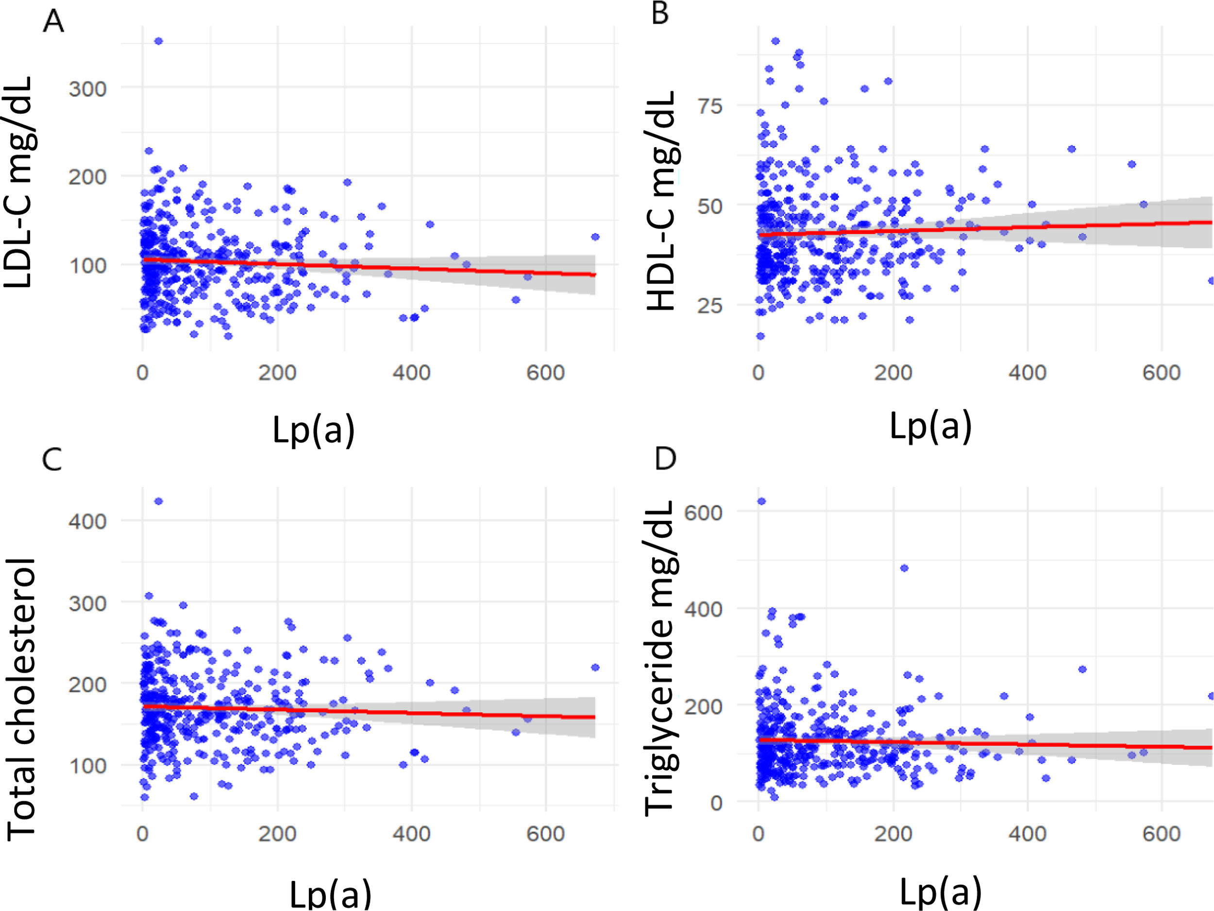
No correlation was found between Lp(a) and LDL-C (p=0.217), HDL-C (p=0.291), total cholesterol (p=0.272), or triglyceride (p=0.905) levels (Table 3 and Figure 1).
Correlation between lipoprotein(a) and other lipid parameters.
| Spearman correlation coefficient (ρ) | p-Valuea | |
|---|---|---|
| Lipoprotein(a) | ||
| Total cholesterol | −0.056 | 0.272 |
| LDL cholesterol | −0.063 | 0.217 |
| HDL cholesterol | 0.054 | 0.291 |
| Triglycerides | −0.006 | 0.905 |
HDL: high-density lipoprotein; LDL: low-density lipoprotein.

Correlation between lipoprotein(a) and other lipid parameters. Panel A, Correlation between Lp(a) and LDL-C levels; Panel B, Correlation between Lp(a) and HDL-levels; Panel C, Correlation between Lp(a) and total cholesterol levels; Panel D, Correlation between Lp(a) and triglyceride levels.
The median age of the patients was 63 years (IQR, 55–73). The age distribution did not differ significantly between the lower and elevated Lp(a) groups (p=0.4) (Table 1).
A total of 25.5% of patients had premature CAD. There were no significant differences in Lp(a) levels between patients with non-premature CAD and those with premature CAD (Table 1).
Traditional cardiovascular risk factorsRegarding the presence of traditional cardiovascular risk factors, 53.4% of patients had arterial hypertension, 49.7% had dyslipidemia, 25.5% had diabetes, 25% were obese, 32.5% were active smokers and 20.1% were previous smokers. There were no differences in the prevalence of traditional cardiovascular risk factors between the elevated Lp(a) and lower Lp(a) groups (Table 1).
Overall, when considering the sum of traditional cardiovascular risk factors, 10.6% did not display traditional cardiovascular risk factors, 23.2% had one traditional cardiovascular risk factor, 28.9% had two traditional cardiovascular risk factors, 23.7% had three traditional cardiovascular risk factors, 10.6% had four traditional cardiovascular risk factors and 2.1% had five traditional cardiovascular risk factors (Supplementary Table 2).
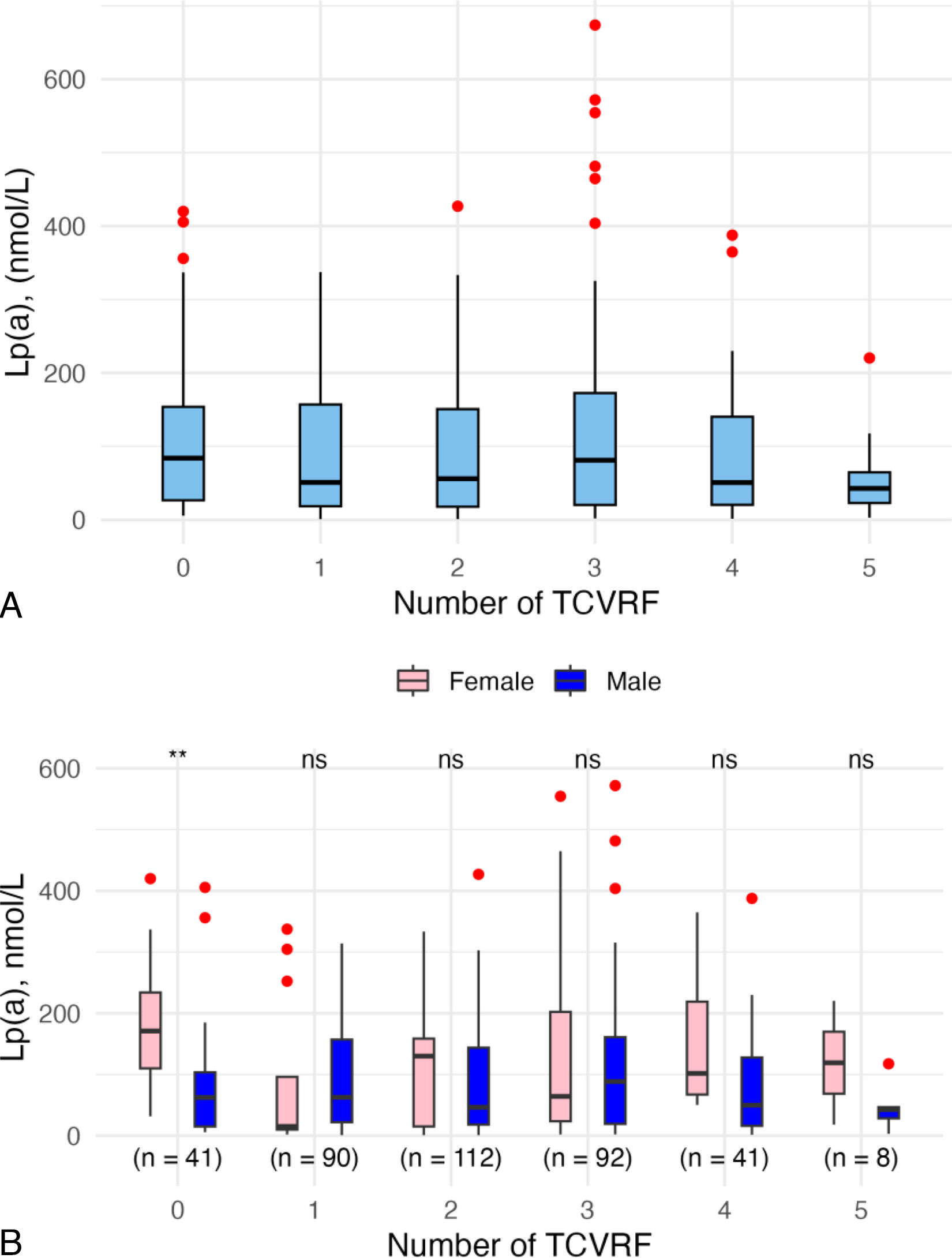
Lp(a) values remained consistent across the different traditional cardiovascular risk factor categories, showing no differences (p=0.7) (Figure 2A and Supplementary Table 2).
Sex
This cohort comprised 19.6% women and 80.4% men. Women tended to have higher Lp(a) levels [47% of female patients with elevated Lp(a) vs. 37% of male patients with elevated Lp(a)] (p-trend=0.014) (Table 1).
When applying the 75 nmol/L cut-off, men represented 84% of patients with Lp(a) levels at or below 75 nmol/L and 77% of those with levels above 75 nmol/L, whereas women accounted for 16% and 23%, respectively. The difference in sex distribution between these two groups was not statistically significant. Considering the 175 nmol/L cut-off, the proportion of men decreased from 83% among patients with Lp(a) levels below 175 nmol/L to 72% among those with levels above this threshold, while the proportion of women increased from 17% to 28%, showing a statistically significant difference (p=0.030) (Table 2).
Women showed higher Lp(a) levels than men in the group of patients with no traditional cardiovascular risk factors (p=0.003), but no significant sex-related differences were noted in patients with one or more traditional cardiovascular risk factors (Figure 2B).
Other comorbiditiesPreviously diagnosed CAD (20.4%) was the most common comorbidity, followed by a family history of CVD (8.8%) and peripheral artery disease (7%) (Table 1).
Patients with a history of CAD had higher Lp(a) values (p-trend=0.003) (Table 1), particularly when using the 175 nmol/L threshold, where a difference was observed between groups (p=0.010) (Table 2).
Patients with a history of atrial fibrillation (AF) or flutter showed a trend toward lower Lp(a) levels (p-trend=0.028) (Table 1). This difference reached significance when patients were stratified using the 75 nmol/L (p=0.016) and 175 nmol/L (p=0.029) thresholds (Table 2).
Pre-admission pharmacological treatmentPatients who had been previously treated with high-intensity statins, either alone or in combination with ezetimibe, showed a tendency toward higher Lp(a) levels (p-trend=0.032 and p-trend=0.014, respectively) (Table 1). When patients were stratified according to the 175 nmol/L Lp(a) cut-off, there were also a number of patients in the higher Lp(a) group treated with high-intensity statins (p=0.045) and ezetimibe (p=0.003) (Table 2).
DiscussionLp(a) is a well-established risk factor for CV events, including ACS. The lack of standardized guidelines for its assessment motivated this study, which aimed to characterize Lp(a) levels in a cohort of patients with ACS and explore potential associations with specific clinical characteristics.
Characterization of Lp(a) levelsFirst, our findings showed that Lp(a) levels were heterogeneously distributed within the population, with no clear associations with most individual characteristics or traditional cardiovascular risk factors studied. These results emphasize the importance of universal screening, as recommended by the EAS and the European Society of Cardiology (ESC), to prevent underestimation of CV risk.10,16 Second, we found that more than one-third of the ACS patients had Lp(a) levels greater than 100 nmol/L, representing a relatively high proportion of individuals at increased CV risk, which is consistent with previous literature.11 Notably, the median Lp(a) level in our study (62.0 nmol/L, IQR 18.4–153.8) appears higher than in the general population (19.6 nmol/L, IQR 7.6–74.8) and in other studies, such as Nissen et al., who reported a median of 42.0 nmol/L (IQR 15.0–155.4) in a large multicentre epidemiological cohort of ASCVD patients from 48 countries.17,18 Similarly, Altmann et al. found median Lp(a) levels of 37.05 nmol/L (Min–Max 0.70–886.00) and 28.70 nmol/L (Min–Max 1.60–626.00) in patients with myocardial infarction (MI) aged <60 years and ≥60 years, respectively.19 Additionally, a recent Portuguese study by Saraiva et al. reported exceptionally high Lp(a) values (median 100.50 nmol/L, IQR 19.30–224.70) in 255 ASCVD patients, with a median age of 57.07 years, who were predominantly men (78.4%).20 These comparisons suggest a potentially higher prevalence of elevated Lp(a) in our cohort, warranting further investigation to better characterize Lp(a) levels in the Portuguese population.
This study also provides important information for ongoing phase III trials of Lp(a)-lowering therapies. We observed that 21.1% of our patients had Lp(a) levels >175 nmol/L, a threshold that may qualify them for Lp(a)-lowering therapies, such as pelacarsen, which is currently being evaluated in the Lp(a) HORIZON trial.21 This highlights a potential opportunity for further CV risk reduction through targeted interventions.
The Lp(a) measurement method remains controversial, primarily due to the structural polymorphisms of Apo(a) which result in isoforms of varying sizes.22 The preferred method for Lp(a) quantification is immunoassay using polyclonal antibodies targeting the highly polymorphic kringle IV type 2 domain of Apo(a) component of Lp(a) particles. Given the significant heterogeneity in Apo(a) isoforms, nmol/L is the recommended unit of measurement over mg/dL to avoid over- or underestimating Lp(a) levels.10,11 A key strength of our study is the adoption of this standardized method, enhancing the reliability and comparability of our findings.
Differences in Lp(a) levels according to sexWomen exhibited higher Lp(a) levels than men, particularly in those who did not exhibit traditional cardiovascular risk factors. This difference may be influenced by age, as previous studies have suggested postmenopausal increases in Lp(a) levels. For instance, Simony et al., using data from the Copenhagen General Population Study, reported that women aged 50–89 years had mean Lp(a) levels 17% higher than men, with a 27% increase post-menopause and a 12% reduction during hormone replacement therapy.23 Given the established link between high Lp(a) levels and premature ASCVD in women, our findings reinforce the need to measure Lp(a) in female patients, particularly those without traditional cardiovascular risk factors.
Association of Lp(a) levels with other comorbidities and traditional cardiovascular risk factorsShiyovich et al. previously demonstrated that among patients <50 years presenting with a first MI and no prior ASCVD, high Lp(a) was associated with an increased risk of ACS, independent of traditional cardiovascular risk factors burden.24 Notably, in our study we did not observe significant differences in lp(a) levels across groups with increasing numbers of traditional cardiovascular risk factors, suggesting that elevated Lp(a) levels may be found in ACS patients regardless of traditional risk factors.
Another key finding was that only 11% of patients with Lp(a) greater than 100 nmol/L had a documented family history of CVD, suggesting that Lp(a) screening should not solely rely on family history. Moreover, only 24% of patients with high Lp(a) levels had previously diagnosed CAD, reinforcing the need for Lp(a) screening in asymptomatic individuals. Previous research has shown that most young patients with elevated Lp(a) had ASCVD risk scores inferior to 7.5% before experiencing their first MI, further questioning the reliance on standard risk calculators when deciding on Lp(a) measurement.25
An unexpected finding was that patients with a history of AF or atrial flutter had lower Lp(a) levels. The relationship between Lp(a) and AF remains unclear. Some authors hypothesize that Lp(a) infiltration into atrial tissue may induce gene expression changes, leading to microcalcifications, cellular damage, and electrical remodeling.26
Given the established causal link between elevated Lp(a) and calcific aortic valve disease, particularly aortic stenosis, future studies should also explore the potential correlation between Lp(a) levels and echocardiographic findings of aortic sclerosis or stenosis in ACS populations.
Pharmacological treatmentPatients who were previously treated with high-intensity statins and ezetimibe tended to have higher Lp(a) levels. A systematic review and meta-analysis of randomized statin trials found that statin therapy did not significantly increase Lp(a) levels nor modify the associated CVD risk.27 Similarly, ezetimibe, whether used alone or in combination with a statin, has been shown to have no impact on plasma Lp(a) levels.28 These findings emphasize the presence of residual CVD risk in patients with high Lp(a) levels despite lipid-lowering therapy. They may also reflect that individuals with higher baseline Lp(a) levels – due to their increased inherent CV risk – are more likely to have experienced a greater number of atherosclerotic events, which in turn leads to more frequent initiation or intensification of lipid-lowering therapy.
Study limitationsThis study faced certain limitations. Firstly, the relatively small sample size, which did not involve a power calculation, might have been inadequate to detect significant relationships between variables. Nonetheless, our main objective was to describe Lp(a) levels in patients with ACS. Second, during the initial implementation of our Lp(a) measurement protocol, adherence was inconsistent, with an increase in compliance observed after the first year of protocol implementation. As a result, only 42% of all ACS admissions during the study period were tested for Lp(a), according to the attending cardiologist's decision. This opportunistic sampling approach may have introduced selection bias, limiting the external validity of our findings and particularly the estimation of the true prevalence of elevated Lp(a) levels. Third, our cohort was predominantly male, which reflects the real-world ACS incidence but limits generalizability to female population. Fourth, data were retrospectively collected from hospital records, making the accuracy of the data dependent on the quality of clinical documentation.
ConclusionLp(a) levels exhibited a heterogeneous distribution in our cohort, with 38.7% of patients presenting with elevated values. Women had higher Lp(a) levels than men, especially among those without traditional cardiovascular risk factors. These findings support routine Lp(a) screening in patients with ACS, regardless of traditional cardiovascular risk factors or other clinical characteristics, with particular attention to women. Future research in larger cohorts should aim to further identify high-risk individuals who may benefit from Lp(a)-lowering therapy.
Ethical statementThis study was approved by the Ethics Committee of Hospital Vila Nova de Gaia/Espinho (document no: 134/2024).
FundingFCT – Portuguese Foundation for Science and Technology, under the scope of the Cardiovascular R&D Center – UnIC (UIDB/00051/2020 and UIDP/00051/2020) and RISE (LA/P/0053/2020).
Conflict of interestNone declared.
The following are the supplementary data to this article: