

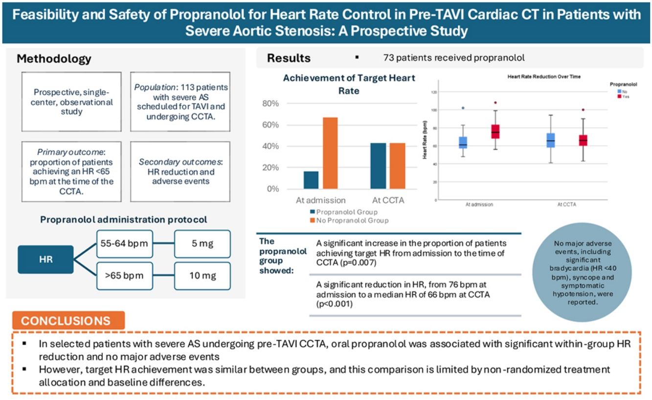
Heart rate (HR) control may improve cardiac computed tomography angiography (CCTA) image quality in preprocedural planning for transcatheter aortic valve implantation (TAVI) in patients with severe aortic stenosis (AS). Beta-blockers, though useful for HR reduction, pose hemodynamic risks in AS patients. Propranolol, a non-selective beta blocker, may offer a safe and effective solution, but its role in this context remains uncertain. The aim of this paper is to assess the feasibility and safety of oral propranolol for HR control in severe AS patients undergoing pre-TAVI CCTA.
MethodsThis prospective, single-center study involved 113 severe AS patients scheduled for pre-TAVI CCTA. Oral propranolol was administered according to baseline HR and individualized clinical assessment: patients with HR 55–64 bpm were eligible for 5 mg, while those with HR >65 bpm were eligible for 10 mg. Propranolol was withheld in patients with lower baseline HR or clinical/hemodynamic features favoring avoidance of beta-blockade. The primary outcome was achieving HR <65 bpm at the time of CCTA. Secondary outcomes included HR reduction and adverse events.
ResultsOf 113 patients, 73 received propranolol and 40 were withheld propranolol. At admission, patients receiving propranolol had higher HR, whereas those not receiving propranolol more frequently had atrial fibrillation, coronary artery disease, and previous beta blocker use. Propranolol group showed significant HR reduction from 76.3±11 bpm to a median HR of 66 bpm (p<0.001). No HR reduction was noted in the non-propranolol group. At CCTA, 43.4% of propranolol patients met the target HR vs. 43.3% of the non-propranolol group. No major adverse events were observed.
ConclusionIn selected patients with severe AS undergoing pre-TAVI CCTA, oral propranolol was associated with significant within-group HR reduction and no major adverse events. However, target HR achievement was similar between groups, and this comparison is limited by non-randomized treatment allocation and baseline differences. These findings support the feasibility and preliminary safety of propranolol, while further studies are needed to establish its efficacy and impact on image quality.
O controlo da frequência cardíaca (FC) é crucial para a obtenção de angiografia por tomografia computorizada cardíaca (CCTA) de elevada qualidade no planeamento pré-procedimento da implantação percutânea da válvula aórtica (TAVI) em doentes com estenose aórtica (EA) grave. Os beta-bloqueadores, embora úteis na redução da FC, podem acarretar riscos hemodinâmicos em doentes com EA. O propranolol, um beta-bloqueante não seletivo, pode oferecer uma solução segura e eficaz, mas o seu papel neste contexto permanece incerto. O objetivo deste estudo foi avaliar a viabilidade e segurança do propranolol oral para controlo da FC em doentes com EA grave submetidos a CCTA pré-TAVI.
MétodosEste estudo prospetivo, unicêntrico, incluiu 113 doentes com EA grave referenciados para CCTA pré-TAVI. O propranolol oral foi administrado de acordo com a FC basal e uma avaliação clínica individualizada: 5mg nos doentes com frequência cardíaca entre 55–64 bpm e 10mg naqueles com FC >65 bpm. O propranolol foi omitido em doentes com FC basal mais baixa ou por razões clínicas/hemodinâmicas que favorecessem evitar beta-bloqueio. O desfecho primário foi a obtenção de uma FC <65 bpm no momento da CCTA. Os desfechos secundários incluíram a redução da FC e a ocorrência de eventos adversos.
ResultadosDos 113 doentes incluídos, 73 receberam propranolol e 40 não o receberam. À admissão, os doentes que receberam propranolol apresentavam uma FC mais elevada, enquanto aqueles que não receberam propranolol apresentavam mais frequentemente fibrilhação auricular, doença arterial coronária e tratamento prévio com beta-bloqueadores. O propranolol associou-se a uma redução significativa da FC, de 76,3 ± 11 bpm à admissão para uma mediana de 66 bpm no momento da CCTA (p<0,001), enquanto não se observou redução significativa no grupo sem propranolol. No momento da CCTA, a FC alvo foi atingida em 43,4% e 43,3% dos doentes com e sem propranolol, respetivamente. Não foram observados eventos adversos major.
ConclusãoEm doentes selecionados com EA grave submetidos a CCTA pré-TAVI, a administração de propranolol oral associou-se a uma redução significativa da FC dentro do grupo tratado e não se associou a eventos adversos major. No entanto, a obtenção da FC alvo no momento da CCTA foi semelhante entre os grupos, sendo esta comparação limitada pela ausência de aleatorização e pelas diferenças basais entre grupos. Estes resultados sugerem a viabilidade e a segurança preliminar do propranolol neste contexto, sendo necessários estudos adicionais para determinar a sua eficácia e o seu impacto na qualidade das imagens.
Aortic stenosis (AS) is the most prevalent valvular heart disease in developed countries.1 Its prevalence is rapidly increasing due to aging population. AS affects 3% of persons older than 65 years and 10% above 80 years old.2 Symptomatic severe aortic stenosis has dismal prognosis and early intervention is recommended.3
Transcatheter aortic valve implantation (TAVI) is an established intervention mode regardless of the surgical risk spectrum, predominantly in elderly patients.1 However, the success of TAVI is highly dependent on meticulous preprocedural planning, where CCTA plays an indispensable role.4 CCTA provides detailed anatomical visualization necessary for accurate valve sizing, assessment of coronary artery involvement, and determination of the optimal fluoroscopic angles for valve deployment.5 CCTA imaging quality may be affected by elevated or irregular HR, particularly for coronary assessment and motion artifact reduction, although strict HR targets may be less critical for aortic annulus sizing alone.5 While betablockers are commonly employed to achieve this target, their use in patients with severe AS is controversial due to the theoretical risk of exacerbating the fixed cardiac output state inherent to AS, potentially leading to hypotension or other hemodynamic instabilities.6 Despite the widespread use of propranolol, a non-selective beta-adrenergic antagonist, for HR control in various clinical settings, its safety and efficacy in patients with severe AS undergoing CCTA have not been evaluated.
ObjectivesThis study aimed to evaluate the feasibility and safety of oral propranolol for HR control in patients with severe AS undergoing pre-TAVI CCTA. We hypothesize that propranolol, despite its non-selective action, may be used in this high-risk population, potentially expanding the options for HR management in CCTA. This study addresses a practical gap in patient preparation for pre-TAVI CCTA.
MethodsStudy designThis was a prospective, observational, single-center study conducted at Unidade Local de Saúde de Coimbra between January 2024 and June 2024. The study enrolled consecutive patients diagnosed with severe AS, defined by an aortic valve area of less than 1.0 cm2 and/or a mean gradient greater than 40 mmHg as assessed by transthoracic echocardiography, and who were selected for TAVI following a Heart Team discussion. Eligible patients were those scheduled for CCTA as part of their pre-TAVI assessment. Exclusion criteria included non-TAVI CCTA, significant bradycardia (HR <40 bpm), hypotension (systolic blood pressure [SBP] <90 mmHg), severe chronic obstructive pulmonary disease, or asthma, and any known contraindications to beta blocker use.
Data collectionBaseline demographics, medical history, and previous medication were obtained through electronic health record review. Heart rate (HR) and systolic blood pressure were recorded at admission and immediately before CCTA. Adverse events were prospectively documented. The study was approved by the local hospital ethics committee (OBS.SF.097/2023).
Propranolol administration protocolOral propranolol was chosen over the more commonly used metoprolol due to its availability at our center. Patients received oral propranolol at least 60 minutes prior to the CCTA scan. The dosage was stratified based on the patients’ baseline HR: those with a resting HR between 55 and 64 bpm received 5 mg, while those with a HR greater than 65 bpm were administered 10 mg. In patients with HR between 40 and 55 bpm, propranolol was systematically withheld. Although patients with HR 55–64 bpm were eligible for propranolol, administration was only performed after individualized clinical assessment. In particular, propranolol was omitted in the presence of borderline hemodynamic parameters—such as HR <50 bpm or systolic blood pressure (SBP) <105 mmHg—or at the discretion of the attending cardiologist.
When patients were positioned on the CT table, one pill of sublingual nitroglycerin (0.5 mg) was given 3–5 minutes before the CT scan to ensure proper visualization of the coronary arteries, based on the patient's blood pressure and the clinician's discretion. HR and blood pressure were measured at baseline (before propranolol administration) and immediately before the CCTA scan (before nitroglycerin administration). Adverse events, including symptomatic bradycardia, hypotension, and bronchospasm, were recorded and managed according to the study standard care. After CCTA, the patient remained on monitoring for 1 hour. Patients were only discharged if hypotension, bradycardia or syncope was not observed. In addition to in-hospital monitoring, a brief one-week follow-up was performed through electronic health record review to identify any emergency department visits or primary care consultations potentially related to adverse effects of propranolol administration (e.g., hypotension, bradycardia, or decompensated heart failure).
Cardiac computed tomography angiography protocolAll CCTA examinations were performed using a 128-slice multidetector computed tomography scanner (SOMATOM Definition AS, Siemens). Imaging parameters were used as follows: slice collimation of 128×0.625 mm, rotation time of 270 ms, tube voltage of 100–120 kV, and tube current of 300–500 mAs depending on patients’ body mass index. The images were acquired using retrospective electrocardiogram triggering at 20–80% phase. The iodinated contrast agent (Ultravist 370, 80–90 mL) was injected into an antecubital vein via an 18G cannula using a dual-syringe technique, at a flow rate of 3.5–5.5 mL/s depending on patients’ body mass index and the tube voltage. Bolus tracking was used with a region of interest placed in the ascending aorta. Images were reconstructed with a slice thickness of 0.8 mm and 0.4-mm increment.
Outcome measuresThe primary outcome was achieving the target HR of <65 bpm at the time of CCTA. Secondary outcomes included the reduction in HR (as a continuous variable) and the incidence of adverse effects related to propranolol administration (SBP <100 mmHg, significant bradycardia [HR <40 bpm], and/or syncope). Image quality was not formally assessed using standardized metrics. Instead, achieving a HR <65 bpm was used as a surrogate marker of optimal image acquisition conditions, consistent with current expert consensus in cardiac CT imaging.5
Sample size calculationWithin-group comparison (propranolol group)The sample size for the within-group comparison (HR at admission vs. HR at the time of the CCTA scan) was based on previous studies showing that oral beta blockers, such as metoprolol, reduced HR to below 65 bpm in 68–86% of patients.7 Using these data, a paired-sample calculation was performed with an expected effect size of 0.26, a one-sided alpha value of 5%, and a power of 80%. The resulting sample size for the propranolol group was 40 patients.
Between-group comparison (propranolol group vs. non-propranolol group)For the between-group comparison, we compared the HR at the time of the CT scan between patients who received propranolol and those who did not because they were already within the target HR. A conservative noninferiority margin of 15% was chosen, as smaller differences are considered clinically irrelevant. The sample size for the between-group comparison required 40 patients in each group to detect significant differences, assuming the same power and alpha values.
Statistical analysisStatistical analyses were performed using SPSS, version 27.0 (APA, MLA, Chicago). Continuous variables, such as HR, were expressed as mean±standard deviation (SD) and compared using the appropriate tests based on distribution. In the within-group analysis (propranolol group), Wilcoxon signed-rank tests were used to compare HR at admission and at the time of the CCTA scan for non-normally distributed data, while paired t-tests were used for normally distributed data. In the between-group analysis (propranolol vs. control), independent t-tests (or Mann–Whitney U tests for non-normally distributed data) were performed to compare the HR at the time of the CCTA scan between the two groups. Categorical variables, such as the incidence of adverse events and the proportion of patients who achieved the target HR, were presented as frequencies and percentages. Comparisons between groups were made using chi-square or Fisher's exact tests, as appropriate. A p-value <0.05 was considered statistically significant for all analyses.
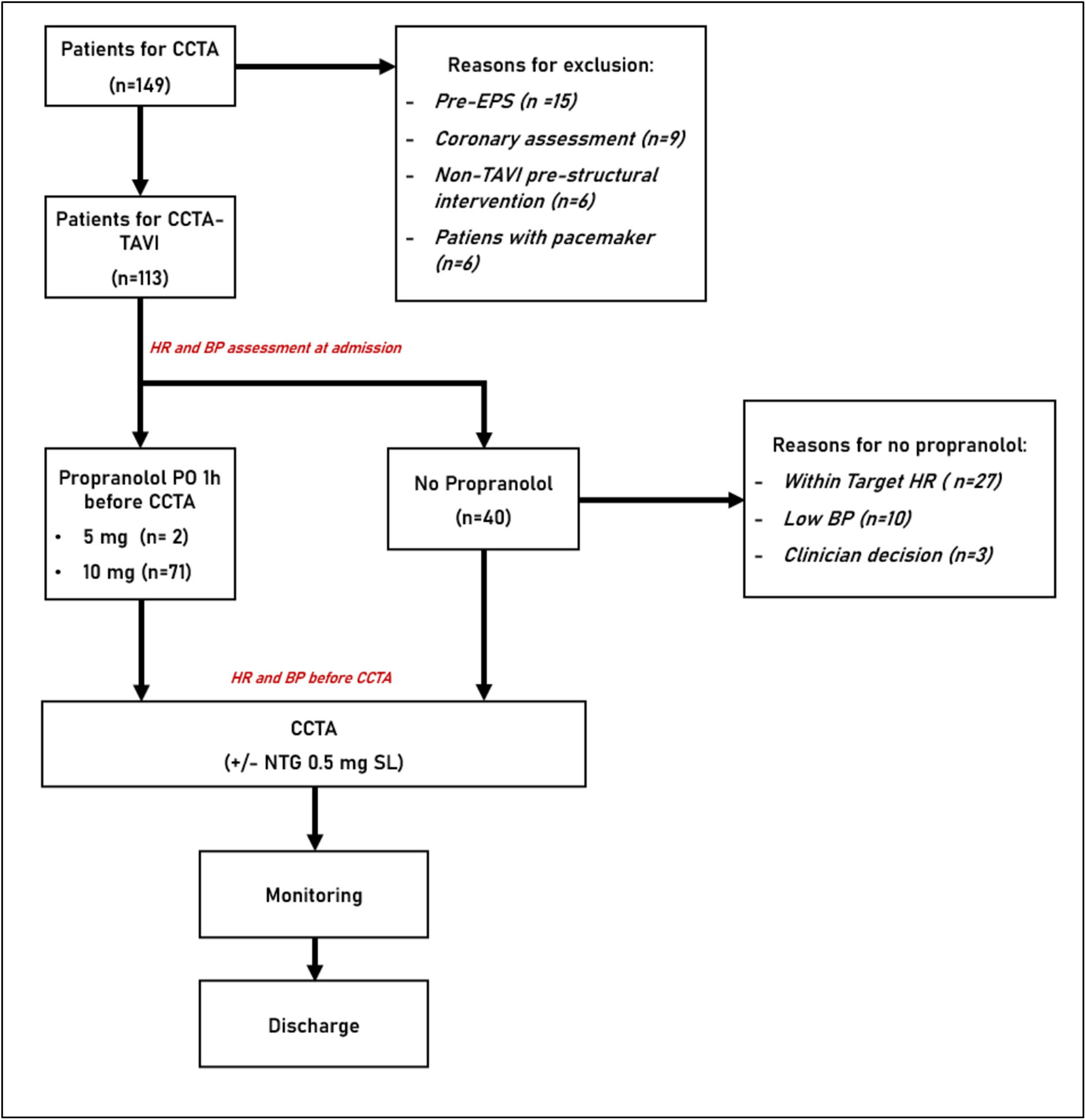
ResultsOf the 149 CCTA performed during the study period, 113 (75,8%) were intended for TAVI planning that complied with our inclusion criteria. 73 patients (64.6%) were administered oral propranolol, and 40 (35,3%) patients did not receive propranolol. Reasons for not administering propranolol are listed in Figure 1. Additionally, 54 (74%) and 22 (55%) patients received sublingual nitrate within the propranolol and non-propranolol group, respectively.
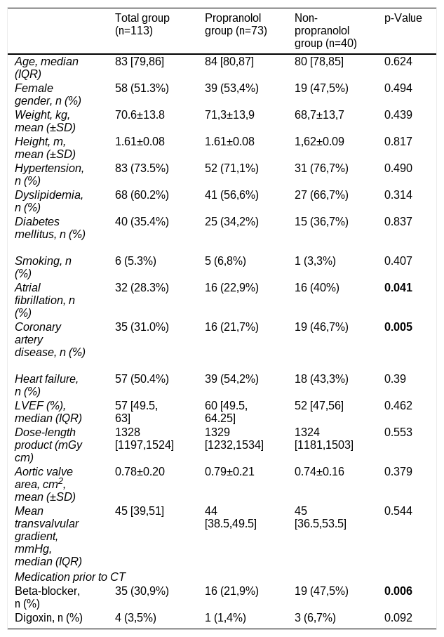
Baseline characteristics were broadly similar between the propranolol and non-propranolol groups, although relevant imbalances were observed. Median age was 84 [80–87] vs. 80 [78–85] years (p=0.624), with 53% vs. 46.7% females (p=0.494). Mean weight (71.3±13.9 kg vs. 68.7±13.7 kg, p=0.439) and height (1.61±0.08 m vs. 1.62±0.09 m, p=0.817) were also comparable. No significant differences were found in hypertension, dyslipidemia, diabetes mellitus or smoking status (p>0.3). However, atrial fibrillation (22.9% vs. 40.0%, p=0.016) and coronary artery disease (21.7% vs. 46.7%, p=0.005) were more prevalent in the non-propranolol group. The severity of aortic stenosis (AS) was comparable between groups. Mean aortic valve area was 0.79±0.21 cm2 in the propranolol group and 0.74±0.16 cm2 in the non-propranolol group (p=0.379). Similarly, mean transvalvular gradient did not differ significantly between groups (propranolol group: 44.0, IQR 11.0 vs. non-propranolol group: 45.0, IQR 17.0; p=0.544). Regarding symptomatic status, the majority of patients were classified as New York Heart Association functional class II (46.0%) or III (38.1%) at baseline, with no statistically significant differences between groups (p=0.737).
Other variables, such as heart failure and left ventricular ejection fraction (LVEF), showed no significant differences. Beta blocker use was higher in the non-propranolol group (47.5% vs. 21.9%, p=0.006). The results are summarized in Table 1. We also assessed baseline conduction abnormalities in the study population. First-degree atrioventricular block was present in 17 patients (15.0%; p=0.556), while intraventricular conduction disturbances were identified in 52 patients (46.0%; p=0.410), including right bundle branch block (n=8), left bundle branch block (n=7), left anterior fascicular block (n=15), right bundle branch block associated with left anterior fascicular block (n=11), and nonspecific intraventricular conduction abnormalities (n=11), with no significant differences between groups. Previous pacemaker implantation was present in 13 patients (11.5%; p=0.303).
Baseline characteristics of the total cohort, propranolol group, and non-propranolol group.
| Total group (n=113) | Propranolol group (n=73) | Non-propranolol group (n=40) | p-Value | |
|---|---|---|---|---|
| Age, median (IQR) | 83 [79,86] | 84 [80,87] | 80 [78,85] | 0.624 |
| Female gender, n (%) | 58 (51.3%) | 39 (53,4%) | 19 (47,5%) | 0.494 |
| Weight, kg, mean (±SD) | 70.6±13.8 | 71,3±13,9 | 68,7±13,7 | 0.439 |
| Height, m, mean (±SD) | 1.61±0.08 | 1.61±0.08 | 1,62±0.09 | 0.817 |
| Hypertension, n (%) | 83 (73.5%) | 52 (71,1%) | 31 (76,7%) | 0.490 |
| Dyslipidemia, n (%) | 68 (60.2%) | 41 (56,6%) | 27 (66,7%) | 0.314 |
| Diabetes mellitus, n (%) | 40 (35.4%) | 25 (34,2%) | 15 (36,7%) | 0.837 |
| Smoking, n (%) | 6 (5.3%) | 5 (6,8%) | 1 (3,3%) | 0.407 |
| Atrial fibrillation, n (%) | 32 (28.3%) | 16 (22,9%) | 16 (40%) | 0.041 |
| Coronary artery disease, n (%) | 35 (31.0%) | 16 (21,7%) | 19 (46,7%) | 0.005 |
| Heart failure, n (%) | 57 (50.4%) | 39 (54,2%) | 18 (43,3%) | 0.39 |
| LVEF (%), median (IQR) | 57 [49.5, 63] | 60 [49.5, 64.25] | 52 [47,56] | 0.462 |
| Dose-length product (mGy cm) | 1328 [1197,1524] | 1329 [1232,1534] | 1324 [1181,1503] | 0.553 |
| Aortic valve area, cm2, mean (±SD) | 0.78±0.20 | 0.79±0.21 | 0.74±0.16 | 0.379 |
| Mean transvalvular gradient, mmHg, median (IQR) | 45 [39,51] | 44 [38.5,49.5] | 45 [36.5,53.5] | 0.544 |
| Medication prior to CT | ||||
| Beta-blocker, n (%) | 35 (30,9%) | 16 (21,9%) | 19 (47,5%) | 0.006 |
| Digoxin, n (%) | 4 (3,5%) | 1 (1,4%) | 3 (6,7%) | 0.092 |
Data are presented as median [interquartile range] for continuous non-normally distributed variables, mean (±SD) for normally distributed continuous variables, and n (%) for categorical variables. Comparisons between the propranolol and non-propranolol groups were made using the Mann–Whitney U test for continuous variables and the Chi-Square or Fisher's Exact Test for categorical variables. A p-value <0.05 was considered statistically significant.
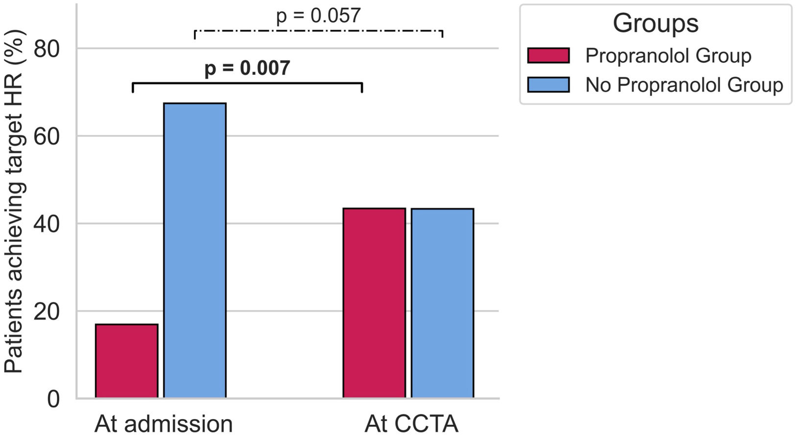
At admission, 83.1% of the propranolol group were above the target HR, whereas 67.4% of the non-propranolol group were already within target (χ2(1)=25.0, p<0.010). At CCTA, 43.4% vs. 43.3% achieved target HR (χ2(1)=0.05, p=0.940). Within-group, the propranolol cohort showed a significant increase in target attainment (χ2(1)=7.37, p=0.007) (Figure 2); the non-propranolol group did not (χ2(1)=3.64, p=0.057).
Proportion of patients in the propranolol and non-propranolol groups who achieved target HR (<65 bpm) at admission and at the time of CCTA. The propranolol group showed a significant increase in the proportion of patients achieving target HR from admission to the time of CCTA (p=0.007), while the non-propranolol group did not show a significant change (p=0.057). CCTA: cardiac computed tomography angiography.
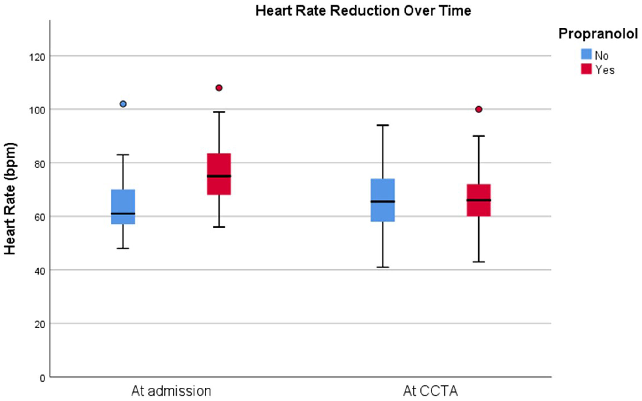
The propranolol group had a mean HR of 76.3±11 bpm at admission and a median HR of 66 bpm (IQR [60,72]) at the time of the CCTA scan, showing a significant reduction. The Wilcoxon Signed-Rank Test confirmed that this reduction was statistically significant (Z=−5.923, p<0.001). The non-propranolol group presented at admission a median HR of 61 bpm (IQR [56,70]) and a median of 65 bpm (IQR [57,75]) at the time of CCTA, with no significant HR reduction (Z=−1.073, p=0.283) (Figure 3).
Box plot showing HR reduction over time for both the propranolol and non-propranolol groups. HR is presented at two-time points: at admission and at the time of CCTA. The propranolol group (red) demonstrates a significant reduction in HR from admission to CCTA (p<0.001), while the non-propranolol group (blue) shows no significant reduction (p=0.283). CCTA: cardiac computed tomography angiography.
At admission, the mean SBP was 136.19±23.86 mmHg in the propranolol group and 135.7±26.28 mmHg in the non-propranolol group, with no significant difference between the groups (p=0.925). At the time of the CCTA scan, the mean SBP in the propranolol group decreased to 133.66±23.7 mmHg, while in the non-propranolol group, it decreased to 129.43±25.9 mmHg, though the difference remained non-significant. Additionally, there was a significant difference in SBP between the nitroglycerin and non-nitroglycerin groups (138.77±20.9 mmHg vs. 117.15±18.3 mmHg, p<0.001).
Adverse outcomesDuring the study period, two patients in the propranolol group developed asymptomatic hypotension (systolic blood pressure <100 mmHg) at the time of the CCTA scan, while two cases were also observed in the non-propranolol group. No instances of significant bradycardia or syncope were documented in either group. No adverse events were identified during the 1-week follow-up period. Review of electronic health records revealed no emergency department readmissions or primary care consultations related to hypotension, bradycardia, syncope, or heart failure decompensation.
DiscussionIn this prospective single-center study of patients with severe AS undergoing pre-TAVI CCTA, oral propranolol was associated with a significant within-group reduction in HR and no major adverse events. However, target HR achievement was similar between patients receiving and not receiving propranolol. This comparison should be interpreted cautiously, since propranolol was not randomly allocated and the non-propranolol group was not a conventional untreated control group. Overall, our study supports the feasibility and preliminary safety of oral propranolol in selected patients, rather than establishing definitive comparative efficacy for target HR achievement.
To our knowledge, this is the first study focusing on the beta blocker use for pre-TAVI CCTA. Furthermore, our study expands the existing body of knowledge by demonstrating that propranolol, despite its non-selective beta-blockade, does not significantly increase the risk of adverse hemodynamic events, such as hypotension or bradycardia, in patients with severe AS. This challenges the conventional caution surrounding non-selective beta blockers in this population and suggests that propranolol may be a viable alternative.8,9 Although strict HR control may be less critical for aortic annulus sizing, lower and more stable HR may be relevant for reducing motion artifacts and improving coronary artery assessment during pre-TAVI CCTA. However, in the present study, coronary interpretability and image quality were not formally assessed using standardized scores. Therefore, the potential benefit of propranolol-related HR reduction on coronary evaluation or overall CCTA diagnostic performance remains speculative and should be addressed in future studies.
Although propranolol was used due to its availability in our center, its pharmacokinetic properties present an equivalent option to metoprolol. Oral propranolol has a relatively short half-life of approximately 3–6 hours,10 while oral metoprolol's half-life ranges from 3 to 7 hours, depending on the formulation (immediate-release vs. extended-release).11 Additionally, propranolol reaches maximum serum concentration 2 hours after oral administration, though this can range from 1 to 4 hours in fasting patients.10 Metoprolol reaches maximum serum concentration 1–2 hours after oral administration.11 Given these properties, propranolol's effects on HR control are sufficient for the brief period required to optimize CCTA imaging. Additionally, receptor selectivity in response to physiological conditions may explain the safety and efficacy of non-selective propranolol in this context.12 When heart rate (HR) is elevated, propranolol primarily exerts its effects on β1-adrenergic receptors located in the atrioventricular (AV) node, effectively reducing HR. Its action on β2-adrenergic receptors in bronchial smooth muscle, which could lead to bronchospasm, is diminished under these conditions. This preferential targeting reduces the likelihood of respiratory side effects, such as bronchospasm, which are a common concern with non-selective beta blockers.12
The use of beta blockers in patients with AS has long been debated due to concerns about their potential to exacerbate left ventricular dysfunction and provoke hemodynamic instability, particularly in the context of severe outflow tract obstruction.13 Historically, this has led to the avoidance of beta blockers in severe AS. However, recent evidence suggests that beta blockers may be both safe and beneficial in managing severe AS, especially in carefully selected patients.14,15 Metoprolol has shown beneficial hemodynamic effects, including reduced heart rate, decreased pressure gradients across the aortic valve, and improved myocardial efficiency.14 Additionally, in patients with moderate to severe asymptomatic AS, beta blockers can help reduce myocardial oxygen demand and alleviate hemodynamic overload without significantly impacting stroke volume.15 Furthermore, data from the OCEAN-TAVI registry demonstrated that preprocedural beta blocker administration was associated with a reduction in in-hospital congestive heart failure and lower cardiovascular mortality in high-risk patients, such as those with prior CABG, peripheral artery disease, elevated BNP, and reduced postprocedural LVEF.16 The study further found no increased risk of procedural complications such as permanent pacemaker implantation, reinforcing the potential safety and efficacy of beta blocker in this population. While these studies focused on other beta blockers than propranolol, and further randomized trials are necessary to solidify these findings, the current evidence supports the cautious use of beta blockers in AS patients.
In our study, sublingual nitroglycerin was administered according to our protocol, without any apparent adverse events. Traditionally, vasodilators were considered contraindicated in patients with severe AS due to concerns that they might precipitate life-threatening hypotension by reducing systemic vascular resistance.13 However, recent studies have demonstrated the beneficial effects of vasodilatation and afterload reduction in patients with severe AS and heart failure.17,18 The UNLOAD Study investigated the use of nitroprusside in intensive care unit patients with depressed heart function (ejection fraction <35%), severe AS (aortic valve area <1.0 cm2), and a low cardiac index (<2.2 L/min/m2). After 24 hours of nitroprusside infusion, the cardiac index increased from 1.6 L/min/m2 to 2.52 L/min/m2 with minimal side effects, demonstrating that nitroprusside can rapidly and markedly improve cardiac function in patients with decompensated heart failure due to severe LV systolic dysfunction and AS.17 This suggests that afterload reduction with arterial vasodilation may improve hemodynamics in patients with severe AS and reduced ejection fraction, though longer-term effects of such therapies require further confirmation. Additionally, recent studies have explored the use of nitroprusside in hypertensive patients with low-gradient severe AS and preserved ejection fraction.18 Eleid et al. demonstrated that nitroprusside infusion improved pulmonary artery pressure, LV end-diastolic pressure, and increased both aortic valve area and mean pressure gradient.18 In our study, patients were positioned lying down on a stretcher prior to the exam and remained in this position for 15–30 minutes post-acquisition, which might mitigate the risk of symptomatic arterial hypotension and ensure hemodynamic stability during the procedure. Albeit not the primary focus of our study and its administration was due to clinician decision, this exploratory result may be studied in future prospective studies.
Study limitationsThis single-center study has several limitations. First, its observational, non-randomized design limits causal inference regarding the efficacy of propranolol. The non-propranolol group should not be interpreted as a conventional control group, since propranolol was withheld mainly in patients with lower baseline HR or clinical/hemodynamic considerations. Accordingly, between-group comparisons for target HR achievement are subject to confounding by indication. Relevant baseline imbalances, including a higher prevalence of atrial fibrillation, coronary artery disease, and previous beta blocker use in the non-propranolol group, may also have influenced HR control and response to treatment.
Second, the relatively small sample size may have limited the detection of rare adverse events, particularly in a high-risk population with severe AS. Third, the lack of comparison with a beta-1 selective agent such as metoprolol prevents definitive conclusions about the relative efficacy and safety of propranolol. Additionally, the proportion of patients achieving the target HR was low in both groups, highlighting the need for improved patient preparation strategies. Fourth, although no adverse events were identified through 1-week electronic health record review, the absence of structured outpatient follow-up or active patient contact may have led to underreporting of mild or delayed symptoms, particularly in this elderly, high-risk population. Fifth, image quality was not assessed using standardized scores (e.g., Likert scale or motion artifacts), although HR <65 bpm is a recognized surrogate marker of optimal acquisition.19 Lastly, the exploratory use of nitroglycerin was based on clinical judgment and was not systematically evaluated.
Future directionsFuture research should focus on conducting multicentre trials with larger and more diverse patient populations to validate these findings and explore the long-term outcomes associated with propranolol use in pre-TAVI CCTA. Comparative studies directly evaluating propranolol against beta-1 selective agents are particularly needed to identify specific patient subgroups that may benefit more from one agent over the other. Moreover, further investigation into the optimal dosing strategies for propranolol, potentially tailored to individual patient characteristics such as body weight, baseline HR, and comorbid conditions, would be valuable.
ConclusionIn selected patients with severe AS undergoing pre-TAVI CCTA, oral propranolol was associated with a significant within-group reduction in HR and no major adverse events. However, target HR achievement at CCTA was similar between patients receiving and not receiving propranolol, a comparison limited by the observational, non-randomized design and baseline differences between groups. These findings support the feasibility and preliminary safety of oral propranolol in this setting.
Declaration of generative AIDuring the preparation of this work, the author(s) used ChatGPT (OpenAI) in order to improve readability and language. After using this tool, the author(s) reviewed and edited the content as needed and take full responsibility for the content of the publication.
Conflicts of interestThe authors have no conflicts of interest to declare.
We extend our sincere gratitude to the nursing staff and radiology technicians whose expertise and dedication made this work possible. Their exceptional patient care, vigilant monitoring, and technical proficiency in imaging were essential to the success of this study. We greatly appreciate their contributions to ensuring both patient safety and high-quality data collection.