
A 57-year-old woman from Mozambique was referred for a cardiology evaluation due to progressive fatigue lasting several months. She denied dyspnea, chest pain, syncope, or peripheral edema. Her medical history was notable for chronic right lower limb lymphedema and bilateral foot dermatophytosis. Physical examination was unremarkable except for right leg edema.
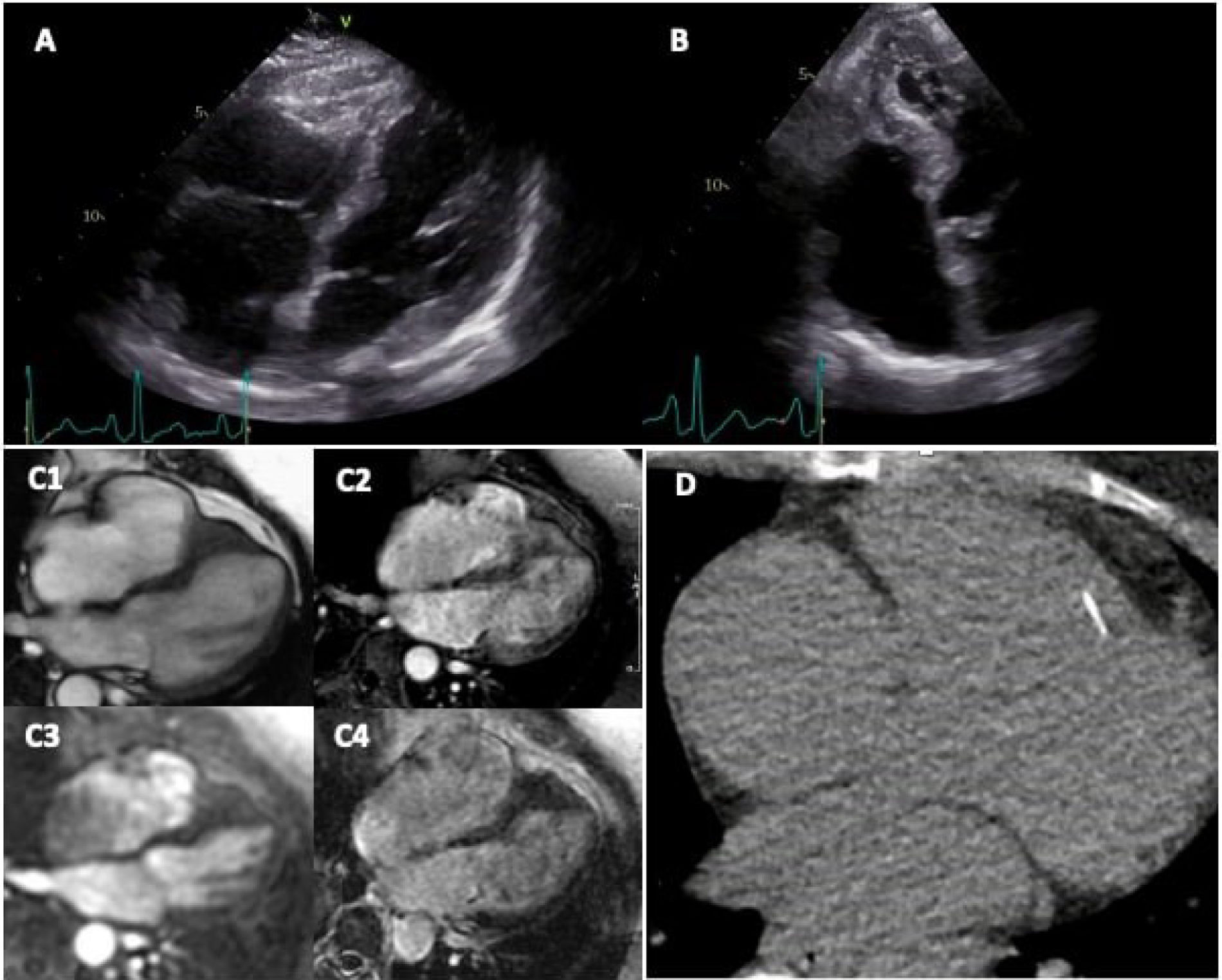
Laboratory evaluation revealed macrocytic anemia, mild leukopenia with a normal eosinophil count, thrombocytopenia, and elevated erythrocyte sedimentation rate (70 mm/h, normal <20), ferritin (1572 ng/mL, normal 30–340), transferrin saturation (85%, normal 15–50), and vitamin B12 (1013 pg/mL, normal 211–911). Immunofixation electrophoresis identified a light chain IgG kappa monoclonal gammopathy. ECG showed sinus rhythm without significant abnormalities. Transthoracic echocardiogram (TTE) demonstrated a mildly dilated right ventricle (RV) with apical obliteration, normal left ventricular size and systolic function, and no other significant abnormalities (Figure 1A and B).

(A and B) TTE, four chamber view showing mildly dilated right ventricle with apical obliteration. (C1) Cardiac MRI, apical right ventricle obliteration. (C2) Cardiac MRI, early gadolinium enhancement, no thrombi. (C3) Cardiac MRI, perfusion, no thrombi. (C4) Cardiac MRI, no late gadolinium enhancement. (D) Cardiac CT calcium score, calcification of the endomyocardial border.
Cardiac MRI confirmed apical RV obliteration (Figure 1C1) with no thrombi (Figure 1C2 and C3), no late gadolinium enhancement (Figure 1C4), and normal native T1, T2*, and extracellular volume (ECV) mappings, findings that do not support endomyocardial fibrosis (EMF), cardiac amyloidosis, or iron overload. However, the CT calcium score revealed a rim of calcification along the RV endomyocardial border (Figure 1D), supporting the hypothesis of EMF.
This case illustrates an early phenotype of isolated RV endomyocardial fibrosis, preceding overt mural thrombosis or extensive fibrosis detectable by cardiac MRI. Cardiac CT calcium scoring proved crucial for the diagnosis. Monoclonal gammopathy could be a bystander, but EMF could also be secondary to a hematological malignancy. Nevertheless, tropical EMF is the most likely cause of the cardiac phenotype. The prevalence of tropical EMF can be as high as 20% in rural areas of Mozambique, hence it should be suspected in symptomatic patients from endemic areas.1,2
Authors’ contributionsAll authors have contributed substantially to the conception, drafting, and revision of the manuscript, and all have approved the final version.
Informed consentThe patient provided written informed consent for publication of the clinical information and imaging findings included in this manuscript. All identifying information has been anonymized to ensure patient confidentiality.
FundingThis article received no external funding.
Conflict of interestThe authors declare no conflict of interest regarding the publication of this manuscript.