Informação da revista
Vol. 35. Núm. 7 - 8.
Páginas 453-454 (Julho - Agosto 2016)
Partilhar
Partilhar
Baixar PDF
Mais opções do artigo
Vol. 35. Núm. 7 - 8.
Páginas 453-454 (Julho - Agosto 2016)
Image in Cardiology
Open Access
Optical coherence tomography images of three different overlapping stents
Sobreposição de três plataformas intracoronárias. Caracterização por tomografía de coerência ótica
Visitas
4484
Rosa Alba Abellás-Sequeiros
Autor para correspondência
albaabellas@gmail.com

Corresponding author.
, Raymundo Ocaranza-Sánchez, Ramiro Trillo-Nouche, José Ramón González-Juanatey
Interventional Cardiology Unit, University Clinical Hospital of Santiago de Compostela, Santiago de Compostela, A Coruña, Spain
Este item recebeu

Under a Creative Commons license
Informação do artigo
Texto Completo
Baixar PDF
Estatísticas
Figuras (3)
Mostrar maisMostrar menos
Material adicional (1)
Texto Completo

A 49-year-old Caucasian woman with prior history of hypertension, diabetes and smoking presented with an inferior ST-elevation myocardial infarction in Killip class I. Ischemia time was four hours. Primary percutaneous coronary intervention (PCI) was performed with thrombus aspiration and direct stenting with a drug-eluting stent (DES). She subsequently underwent PCI with bioresorbable vascular scaffolds (BVS) in the circumflex artery (Cx). After deployment of the BVS a coronary perforation was seen at the distal edge of the device. A stent graft was implanted overlapping the edge of the BVS. The perforation was resolved, nevertheless the distal lesion on the CX artery needed to be revascularized. As another BVS did not cross the proximal BVS, we implanted another metal DES overlapping the stent graft. Optical coherence tomography (OCT) images confirmed right stent expansion and apposition.

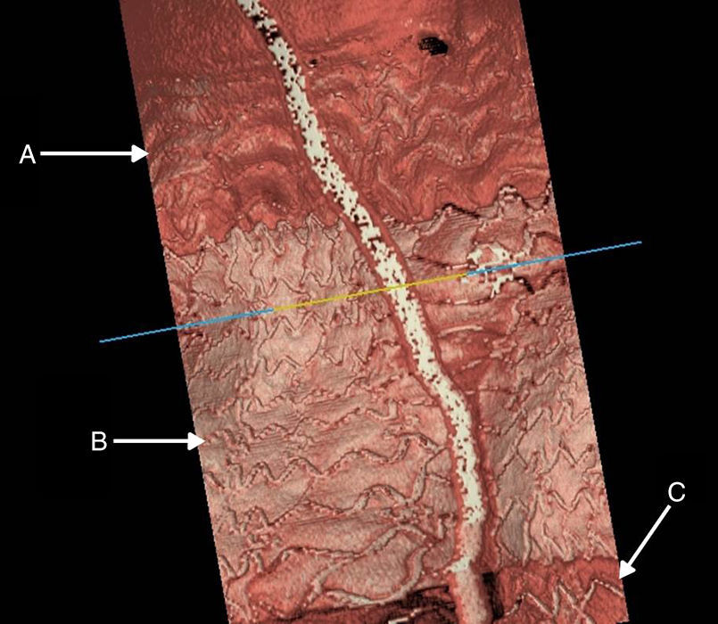
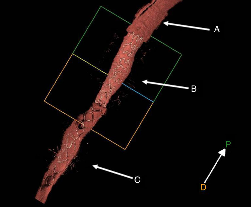
We report an example of successful treatment of a coronary intervention complication with three different kinds of stent. Currently BVS are much less navigable than metal DES; this case is a good example on this characteristic. OCT images show the interface between the resorbable scaffold, the polymer layer of the stent graft and the metal strut of the distal DES (Figures 1 and 2). Figure 3 shows the stent graft overlapping the BVS.

Figure 1.

(A) Bioresorbable strut; (B) polymer stent graft strut; (C) metal strut of the drug-eluting stent.

(0,12MB).
Figure 2.

(A) Bioresorbable strut; (B) polymer stent graft strut; (C) metal strut of the drug-eluting stent.

(0,06MB).
Figure 3.

Optical coherence tomography image showing bioresorbable vascular scaffold struts and distal overlapping of metal struts of the stent graft.

(0,08MB).
Ethical disclosuresProtection of human and animal subjects

The authors declare that no experiments were performed on humans or animals for this study.

Confidentiality of data

The authors declare that no patient data appear in this article.

Right to privacy and informed consent

The authors declare that no patient data appear in this article.

Conflicts of interest

The authors have no conflicts of interest to declare.

Copyright © 2016. Sociedade Portuguesa de Cardiologia
Idiomas
Revista Portuguesa de Cardiologia
Opções de artigo
Ferramentas
Material Suplementar
en pt

Are you a health professional able to prescribe or dispense drugs?

Você é um profissional de saúde habilitado a prescrever ou dispensar medicamentos

Ao assinalar que é «Profissional de Saúde», declara conhecer e aceitar que a responsável pelo tratamento dos dados pessoais dos utilizadores da página de internet da Revista Portuguesa de Cardiologia (RPC), é esta entidade, com sede no Campo Grande, n.º 28, 13.º, 1700-093 Lisboa, com os telefones 217 970 685 e 217 817 630, fax 217 931 095 e com o endereço de correio eletrónico revista@spc.pt. Declaro para todos os fins, que assumo inteira responsabilidade pela veracidade e exatidão da afirmação aqui fornecida.