Informação da revista
Vol. 32. Núm. 11.
Páginas 941-945 (Novembro 2013)
Partilhar
Partilhar
Baixar PDF
Mais opções do artigo
Vol. 32. Núm. 11.
Páginas 941-945 (Novembro 2013)
Image in Cardiology
Open Access
Cardiac amyloidosis: Diagnosis using delayed enhancement cardiac magnetic resonance imaging sequences
Amiloidose cardíaca: diagnóstico através de sequências de realce tardio com o uso de ressonância magnética cardíaca
Visitas
12489
Dinis Mesquitaa,
Autor para correspondência
dinis.mesquita@gmail.com

Corresponding author.
, Carla Nobreb, Boban Thomasc, Nuno Jalles Tavaresc
a Cardiology Department, Centro Hospitalar Barreiro Montijo, Barreiro, Portugal
b Internal Medicine Department, Centro Hospitalar Barreiro Montijo, Barreiro, Portugal
c Caselas MR Centre, Lisbon, Portugal
Este item recebeu

Under a Creative Commons license
Informação do artigo
Texto Completo
Bibliografia
Baixar PDF
Estatísticas
Figuras (7)
Mostrar maisMostrar menos
Texto Completo

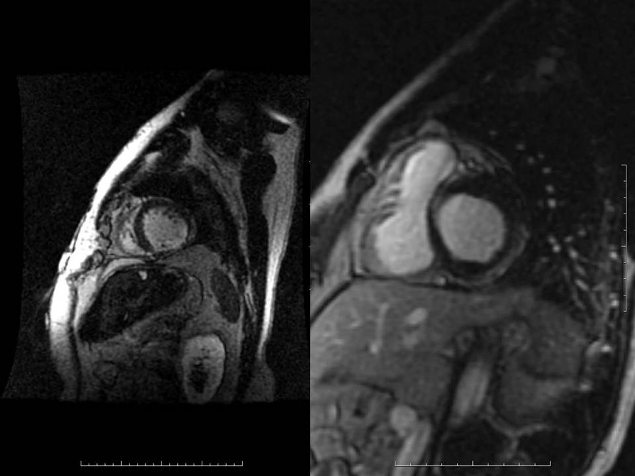
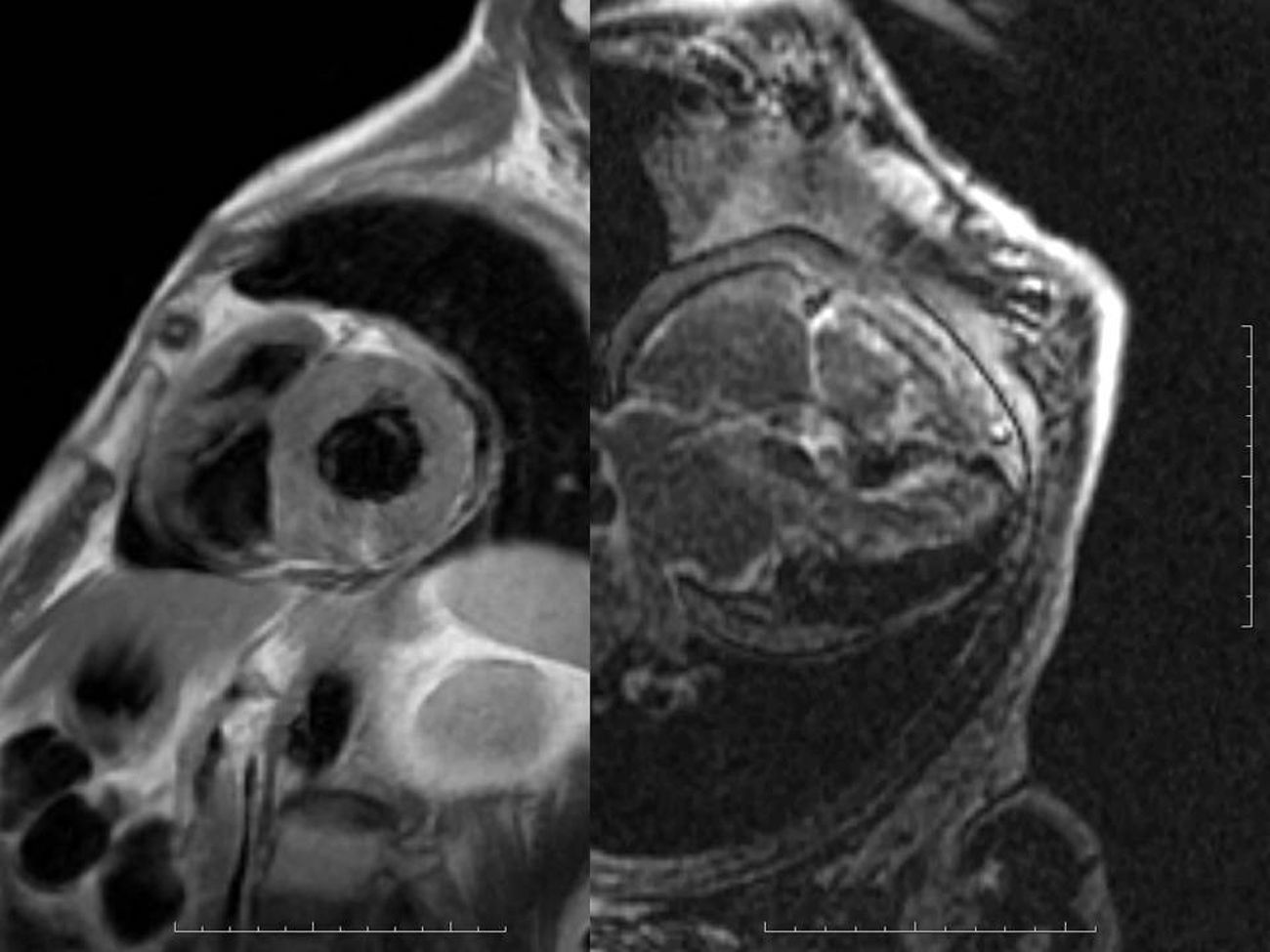
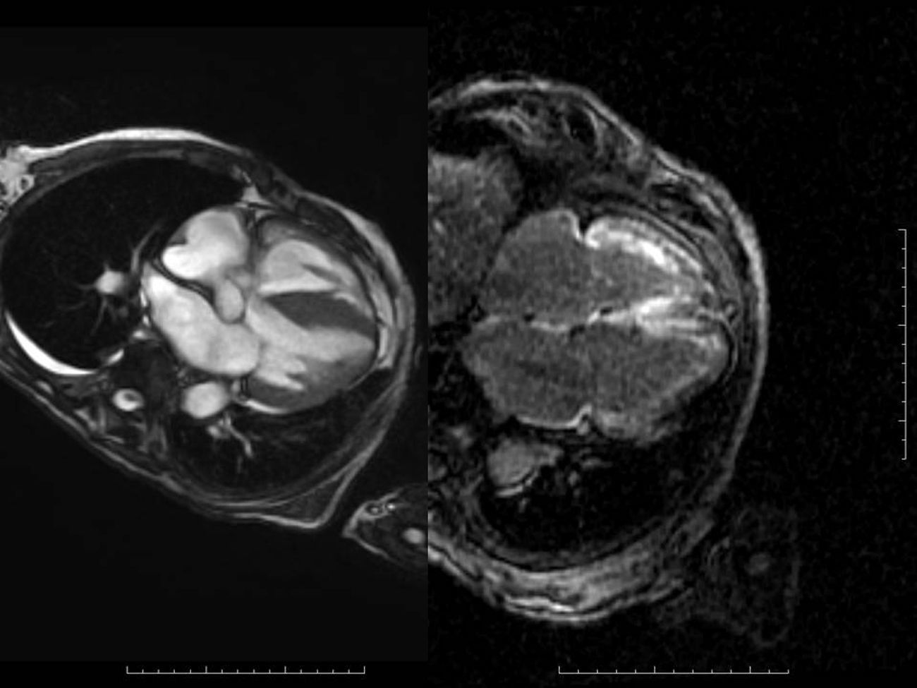
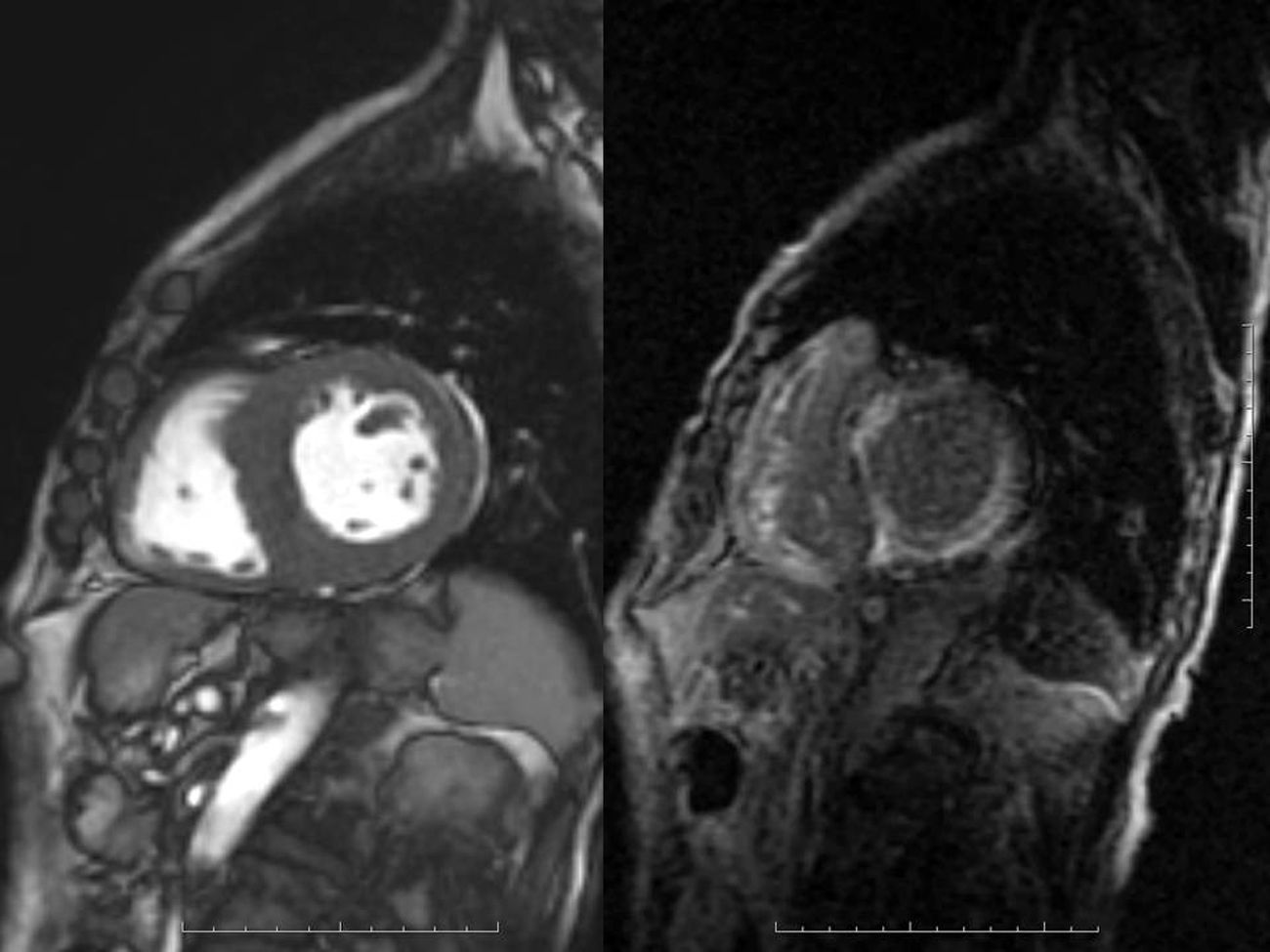
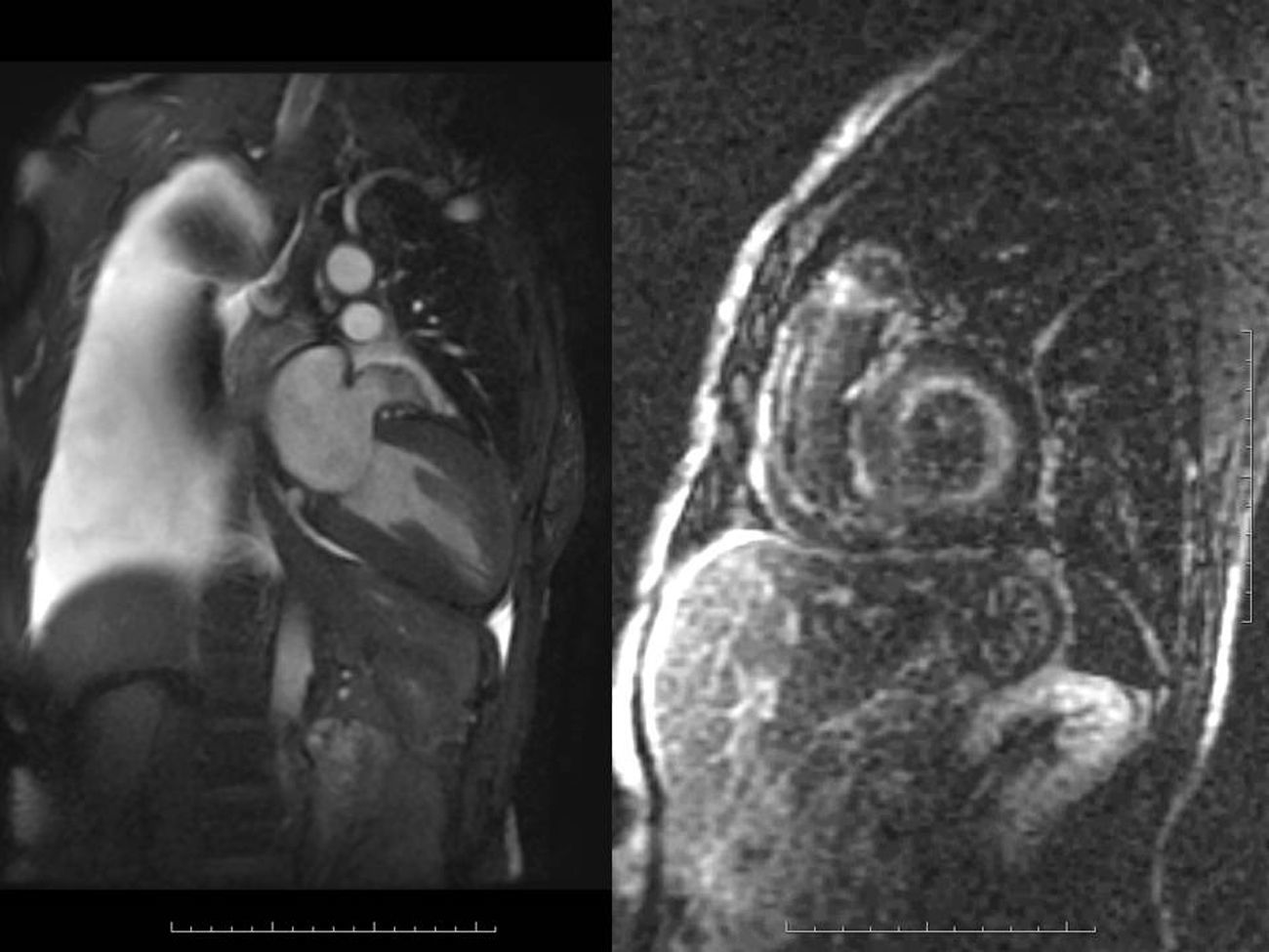
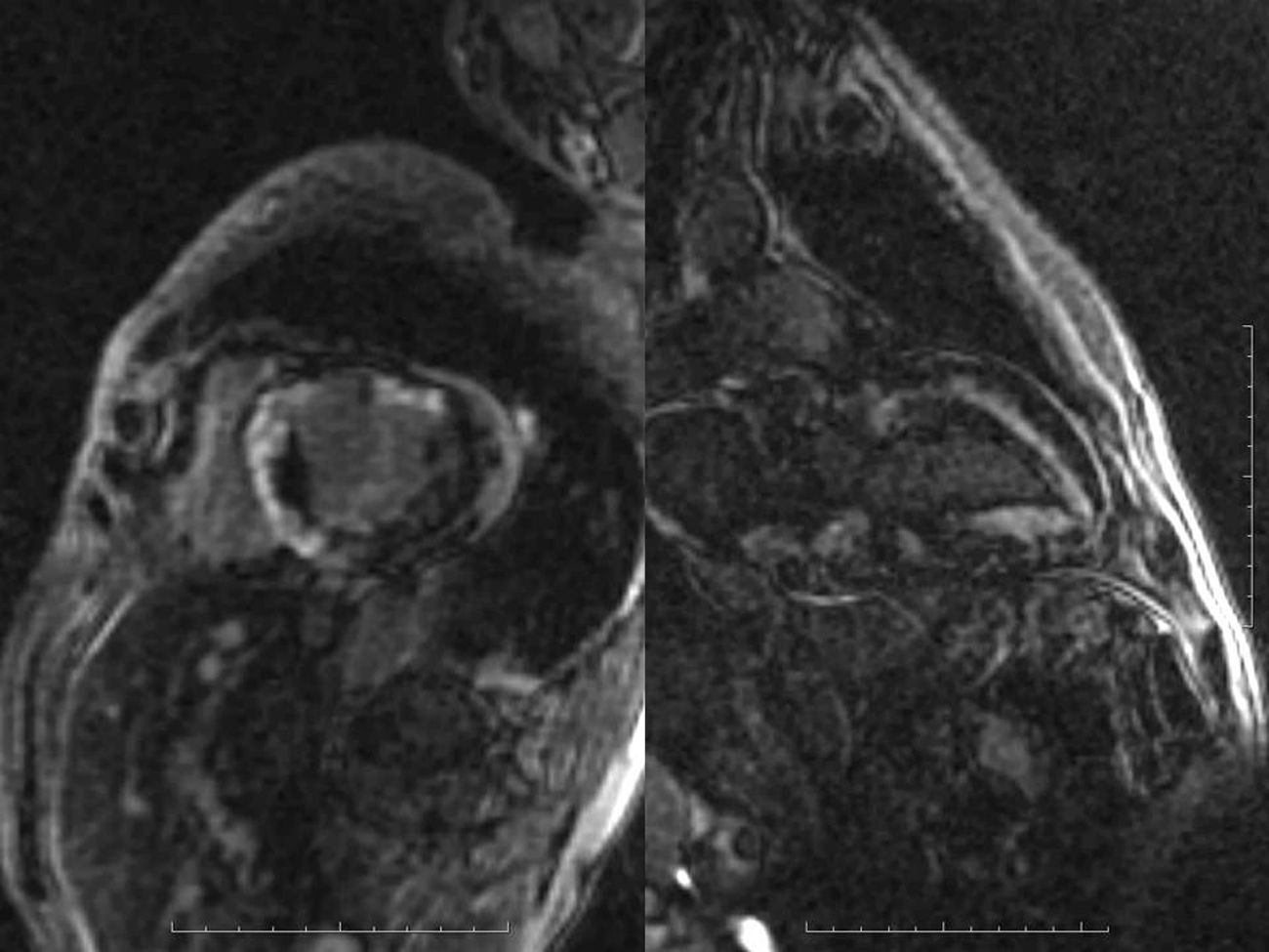
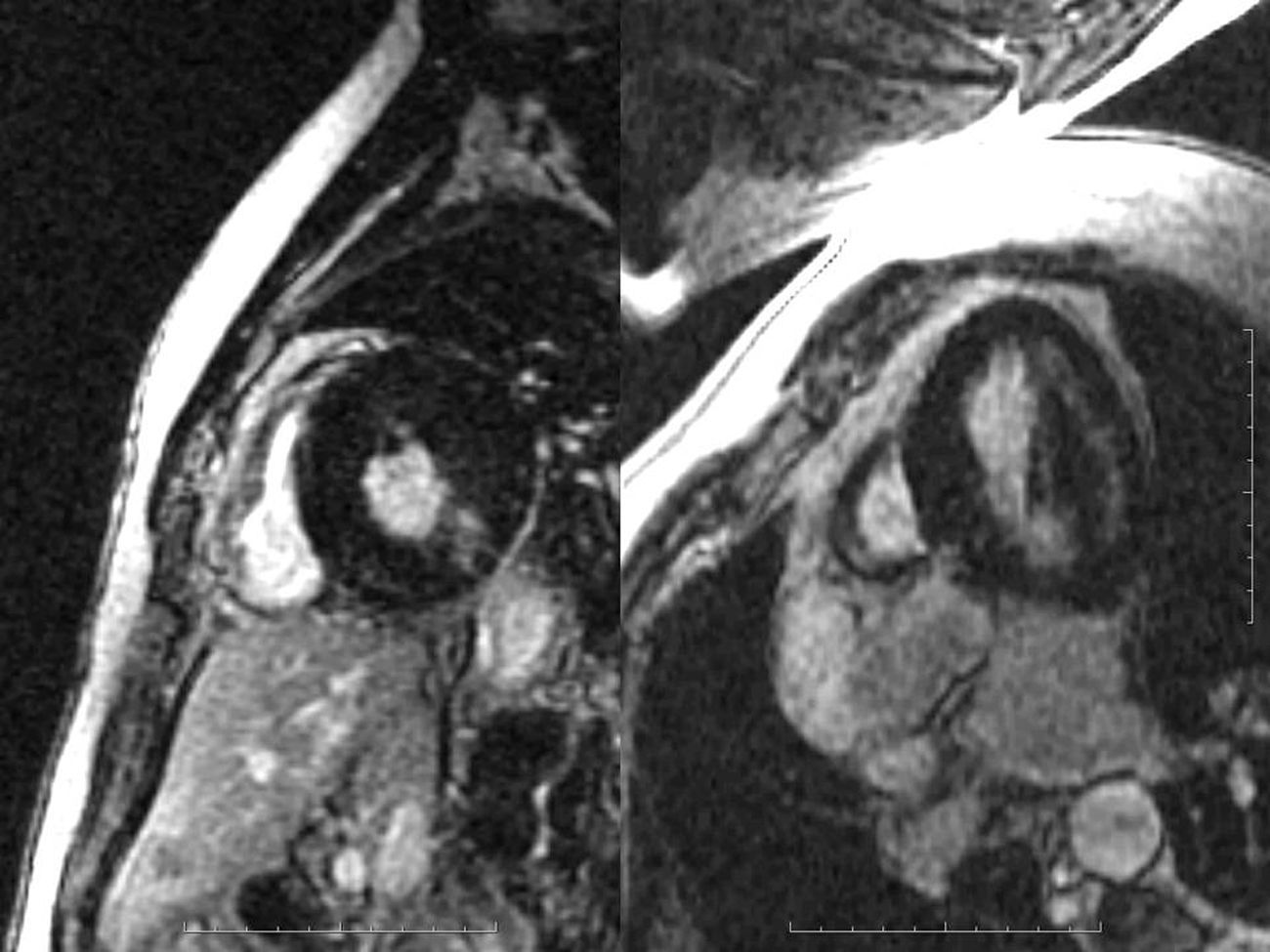
Late gadolinium enhancement (LGE) cardiac magnetic resonance imaging (MRI) is very useful in distinguishing between myocardial infarction, in which the enhancement is typically subendocardial, and myocarditis, in which it is subepicardial (Figure 1). In addition, various patterns have been described with different cardiomyopathies, in some cases enabling a specific diagnosis without invasive workup and risk stratification.1 Cardiac amyloidosis was previously thought to be present only when systemic amyloidosis was patently manifest. This has proved not to be the case, with cardiac MRI detecting increasing numbers of cases in patients with diastolic heart failure in whom cardiac involvement may be the first or sole manifestation. The pattern of LGE commonly found in cardiac amyloidosis is a global subendocardial enhancement with different contrast kinetics, the ventricular cavities showing no signal at all (Figures 2–6) compared to myocarditis and acute myocardial infarction, in which there is an intracavitary gray signal (Figure 1). Of 10 patients referred after echocardiography raised the suspicion of a cardiomyopathy, five were diagnosed with cardiac amyloidosis, two had images not suggestive of amyloidosis and were subsequently found to have Fabry's disease (Figure 7), and the other three probably had concentric left ventricular hypertrophy due to hypertension.

Figure 1.

Left: the inferolateral wall with a transmural infarct with obvious wall thinning, while the left ventricular cavity shows a gray signal due to contrast admixed with blood. Right: a patient with acute myocarditis with subepicardial lateral wall enhancement.

(0,11MB).
Figure 2.

The panel on the left is a T1-weighted image with wall thickening. Beside is the LGE image showing global subendocardial enhancement with no cavitary signal. Both ventricles are involved.

(0,12MB).
Figure 3.

Biventricular global subendocardial enhancement in another patient. Note the biatrial enlargement typical of restrictive cardiomyopathies.

(0,1MB).
Figure 4.

Short-axis views of a patient with extensive global subendocardial involvement.

(0,11MB).
Figure 5.

A patient with decompensated congestive heart failure as evidenced by massive pleural effusion (left) had very good LV systolic function with concentric hypertrophy, while the LGE sequence (right) was diagnostic for amyloidosis.

(0,12MB).
Figure 6.

The patchy involvement seen in this patient can also be seen in amyloidosis. The decreased signal in the ventricular cavity provides a clue.

(0,12MB).
Figure 7.

A patient initially thought to have amyloidosis is found to have no subendocardial LGE. The signal is also present in the cavity. This patient has mid-lateral wall involvement and was later diagnosed with Fabry's disease.

(0,15MB).
Ethical disclosuresProtection of human and animal subjects

The authors declare that no experiments were performed on humans or animals for this study.

Confidentiality of data

The authors declare that no patient data appear in this article.

Right to privacy and informed consent

The authors declare that no patient data appear in this article.

Conflicts of interest

The authors have no conflicts of interest to declare.

Reference
[1]
H. Vogelsberg, H. Mahrholdt, C.C. Deluigi, et al.
Cardiovascular magnetic resonance in clinically suspected cardiac amyloidosis. Noninvasive imaging compared to endomyocardial biopsy.
J Am Coll Cardiol, 51 (2008), pp. 1022-1030
Copyright © 2013. Sociedade Portuguesa de Cardiologia
Idiomas
Revista Portuguesa de Cardiologia
Opções de artigo
Ferramentas
en pt

Are you a health professional able to prescribe or dispense drugs?

Você é um profissional de saúde habilitado a prescrever ou dispensar medicamentos

Ao assinalar que é «Profissional de Saúde», declara conhecer e aceitar que a responsável pelo tratamento dos dados pessoais dos utilizadores da página de internet da Revista Portuguesa de Cardiologia (RPC), é esta entidade, com sede no Campo Grande, n.º 28, 13.º, 1700-093 Lisboa, com os telefones 217 970 685 e 217 817 630, fax 217 931 095 e com o endereço de correio eletrónico revista@spc.pt. Declaro para todos os fins, que assumo inteira responsabilidade pela veracidade e exatidão da afirmação aqui fornecida.