
Even with recent advances in computed tomography (CT) imaging of coronary stents, blooming is still an important limitation in the assessment of stents. Recently, the introduction of bioabsorbable radiolucent scaffolds has allowed almost unrestricted imaging of the stent lumen.
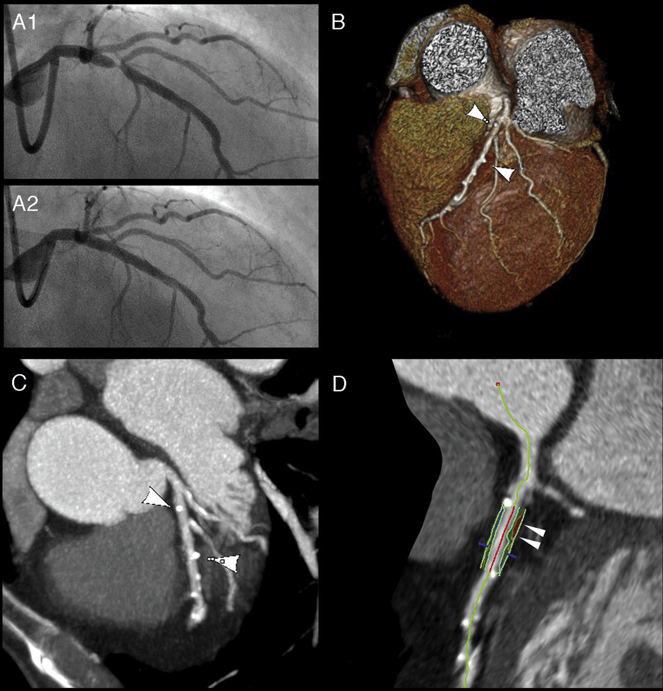
A 68-year-old man with a history of coronary artery disease was admitted with non-ST-elevation myocardial infarction. Coronary angiography revealed 95% stenosis of the proximal left anterior descending artery (Figure 1A1). The patient underwent percutaneous coronary intervention with implantation of a 3.5 mm×18 mm bioabsorbable everolimus-eluting poly(lactic acid) scaffold (Abbott Vascular, Santa Clara, CA) (Figure 1A2). As part of a prospective study, 64-slice CT angiography (SOMATOM Sensation 64 scanner, Siemens, Forchheim, Germany) was performed at the first month follow-up. Radiopaque indicators (arrows) at the edges of the scaffold were the only discernible traces on volume-rendered imaging (Figure 1B) and in maximum intensity projection reconstructions (Figure 1C).
 and angiographic outcome after placement of a bioabsorbable scaffold (A2). Multislice computed tomography angiography: volume rendering (B) and maximum intensity projection reconstructions (C), revealing the radiopaque indicators (arrows) at the edges of the scaffold. Virtual histology technique showing the non-calcified atherosclerotic plaque (arrows) excluded by the stent (D).")
Coronary angiography, showing significant left anterior descending stenosis (A1) and angiographic outcome after placement of a bioabsorbable scaffold (A2). Multislice computed tomography angiography: volume rendering (B) and maximum intensity projection reconstructions (C), revealing the radiopaque indicators (arrows) at the edges of the scaffold. Virtual histology technique showing the non-calcified atherosclerotic plaque (arrows) excluded by the stent (D).
Virtual histology (Aquarius workstation, TeraRecon Inc., San Mateo, CA) showed the non-calcified atherosclerotic plaque (arrows) excluded by the stent (Figure 1D). Cross-sectional in-stent lumen areas were measured, with a minimal lumen area of 5.54 mm2. No significant lumen loss was detected.
This case illustrates the new opportunities generated by the use of novel bioabsorbable scaffolds. CT angiography indications may expand to the follow-up of smaller stented vessels and new insights into the process of atherosclerotic healing after stenting can be expected.
Ethical disclosuresProtection of human and animal subjectsThe authors declare that no experiments were performed on humans or animals for this study.
Confidentiality of dataThe authors declare that they have followed the protocols of their work center on the publication of patient data.
Right to privacy and informed consentThe authors have obtained the written informed consent of the patients or subjects mentioned in the article. The corresponding author is in possession of this document.
Conflicts of interestThe authors have no conflicts of interest to declare.
