

Pseudoaneurysms are a serious complication of myocardial infarction but can be asymptomatic and only detected by chance in patients with previous subclinical events.
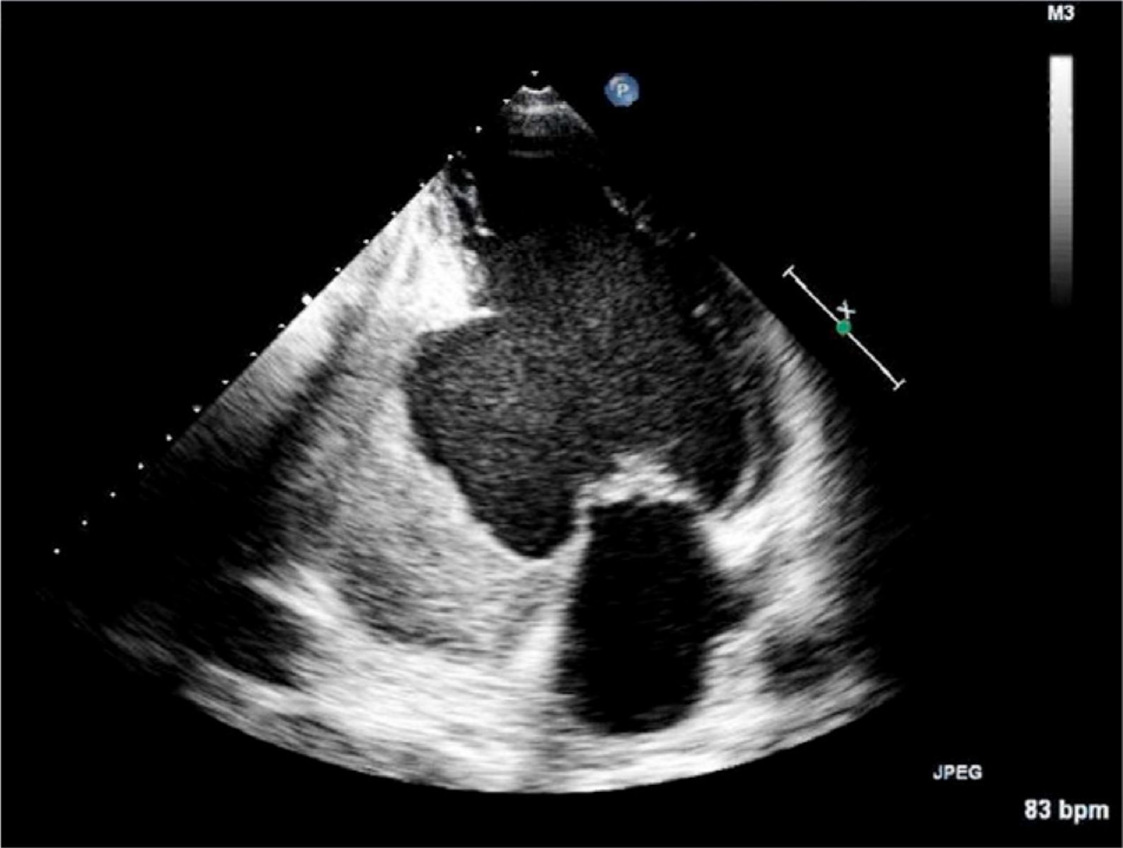
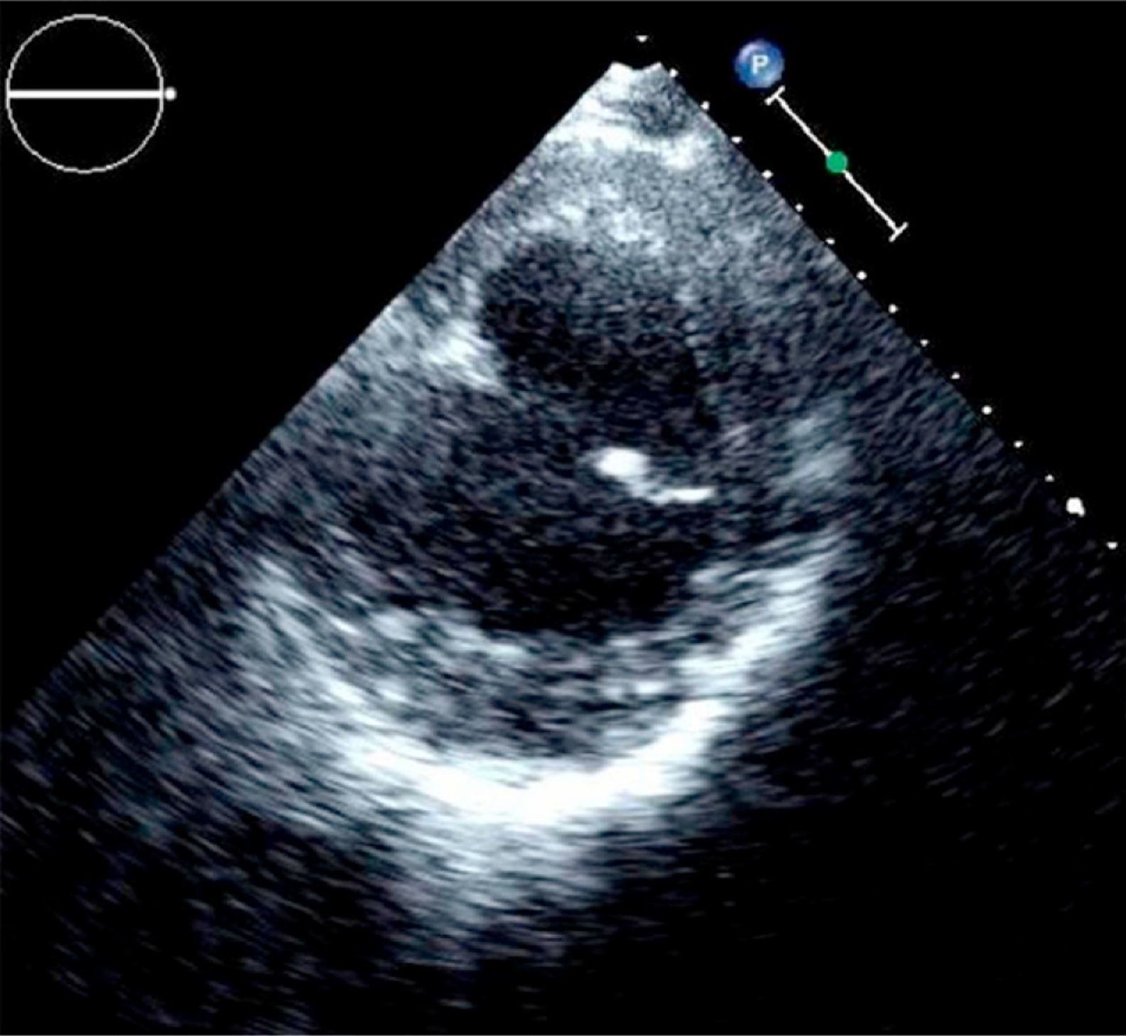
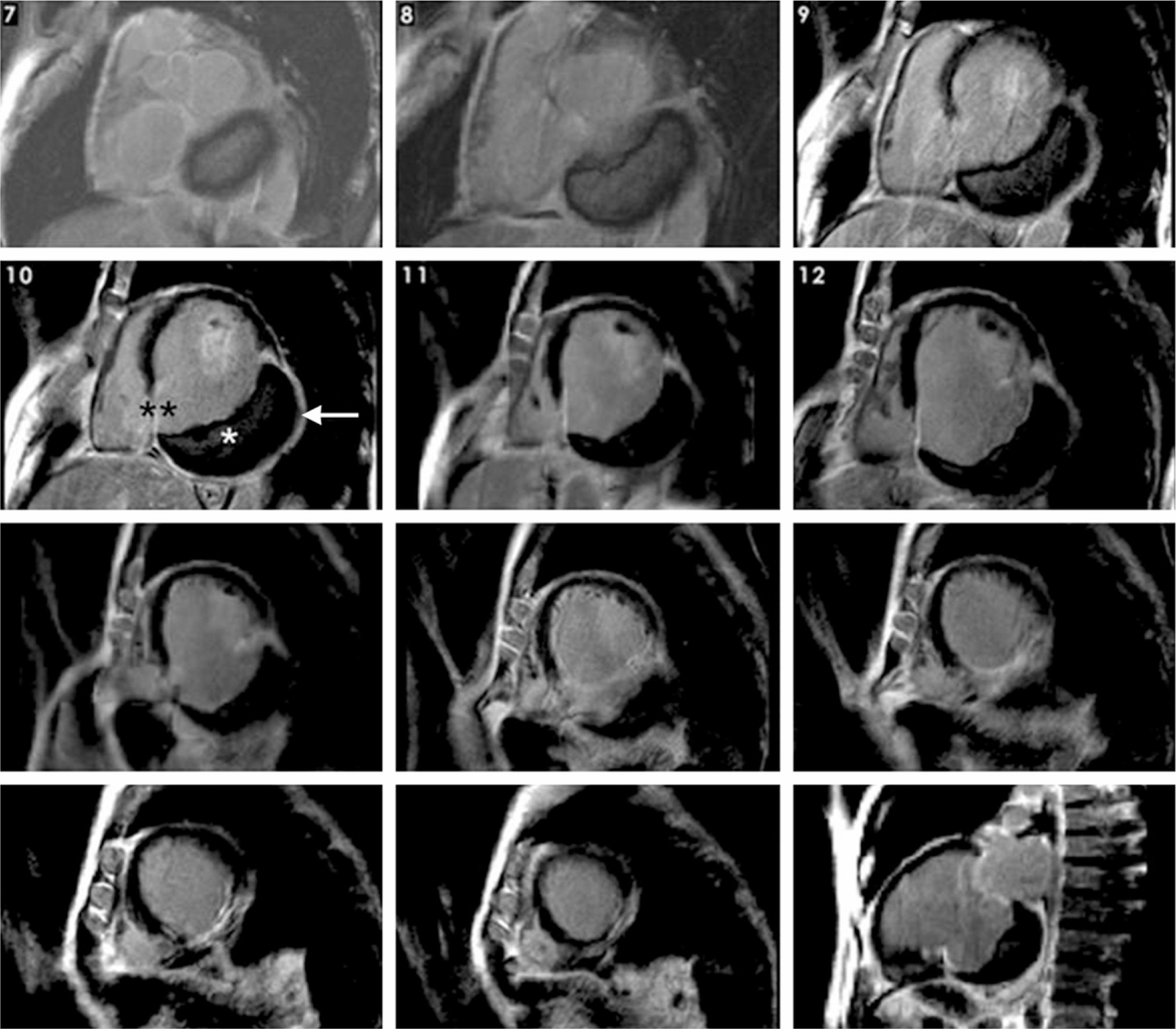
The images presented are of a 62-year-old man, a smoker, with no history of heart disease, admitted to our hospital for community-acquired pneumonia. The chest X-ray on admission showed cardiomegaly and the electrocardiogram showed Q waves in the inferior leads. Transthoracic echocardiography revealed mild left chamber dilatation and severe left ventricular systolic dysfunction, with an aneurysmal sac, 8cm in diameter, involving the basal portion of the posterior, inferior and lateral walls and containing a thrombus (Figures 1 and 2). Since unequivocal differential diagnosis between aneurysm and pseudoaneurysm was not possible with this technique, magnetic resonance imaging was performed, which revealed an old extensive inferior infarction, extending to the adjacent portion of the right ventricle, with inferior myocardial wall rupture. A diagnosis was thus established of pseudoaneurysm in the inferior and posterior walls, containing an organized thrombus (Figures 3 and 4). Cardiac catheterization showed single-vessel disease with chronic occlusion of the mid segment of the right coronary artery. Surgical repair was proposed but the patient refused. He remained clinically stable in NYHA class II for 10 months after discharge, but was then lost to follow-up.


, showing transmural infarction of the inferior basal and mid segments. The basal inferior segment of the left ventricular wall consists of a giant thrombus (*), infarcted myocardium (**) and pericardium (arrow), confirming a pseudoaneurysm.")
Delayed enhancement cardiac magnetic resonance images, in short-axis view from the base to the apex and two-chamber view (right bottom corner), showing transmural infarction of the inferior basal and mid segments. The basal inferior segment of the left ventricular wall consists of a giant thrombus (*), infarcted myocardium (**) and pericardium (arrow), confirming a pseudoaneurysm.
This case illustrates the value of complementary imaging techniques for correct diagnosis of a complication of an extensive asymptomatic infarction.
Ethical disclosuresProtection of human and animal subjectsThe authors declare that no experiments were performed on humans or animals for this investigation.
Confidentiality of dataThe authors declare that they have followed the protocols of their work center on the publication of patient data and that all the patients included in the study have received sufficient information and have given their informed consent in writing to participate in the study.
Right to privacy and informed consentThe authors declare that no patient data appears in this article.
Conflicts of interestThe authors have no conflicts of interest to declare.
Please cite this article as: Antunes N, et al. Pseudoaneurisma gigante do ventrículo esquerdo- catástrofe silenciosa. Rev Port Cardiol. 2012. doi:10.1016/j.repc.2012.05.010.
