

We describe the case of a 64-year-old woman with persistent atrial fibrillation (more than one year's duration) referred for ablation. Three-dimensional electroanatomical mapping of the left atrium was performed using the Ensite NavX™ system (St. Jude Medical, Minneapolis, MN) and a Constellation™ 64-pole basket diagnostic catheter (Boston Scientific, Natick, MA). A stable complex fragmented atrial electrogram (CFAE) was identified in 5-min time-frame windows using novel proprietary software (Figure 1). A localized radiofrequency application over the CFAE near the left pulmonary vein ostia modified electrical activity in the posterior wall of the left atrium, increasing cycle length until interruption (Figures 2–4). This case highlights the importance of finding areas that are critical in maintaining electrical fibrillatory activity. A precise ablation target was identified (stable CFAE).
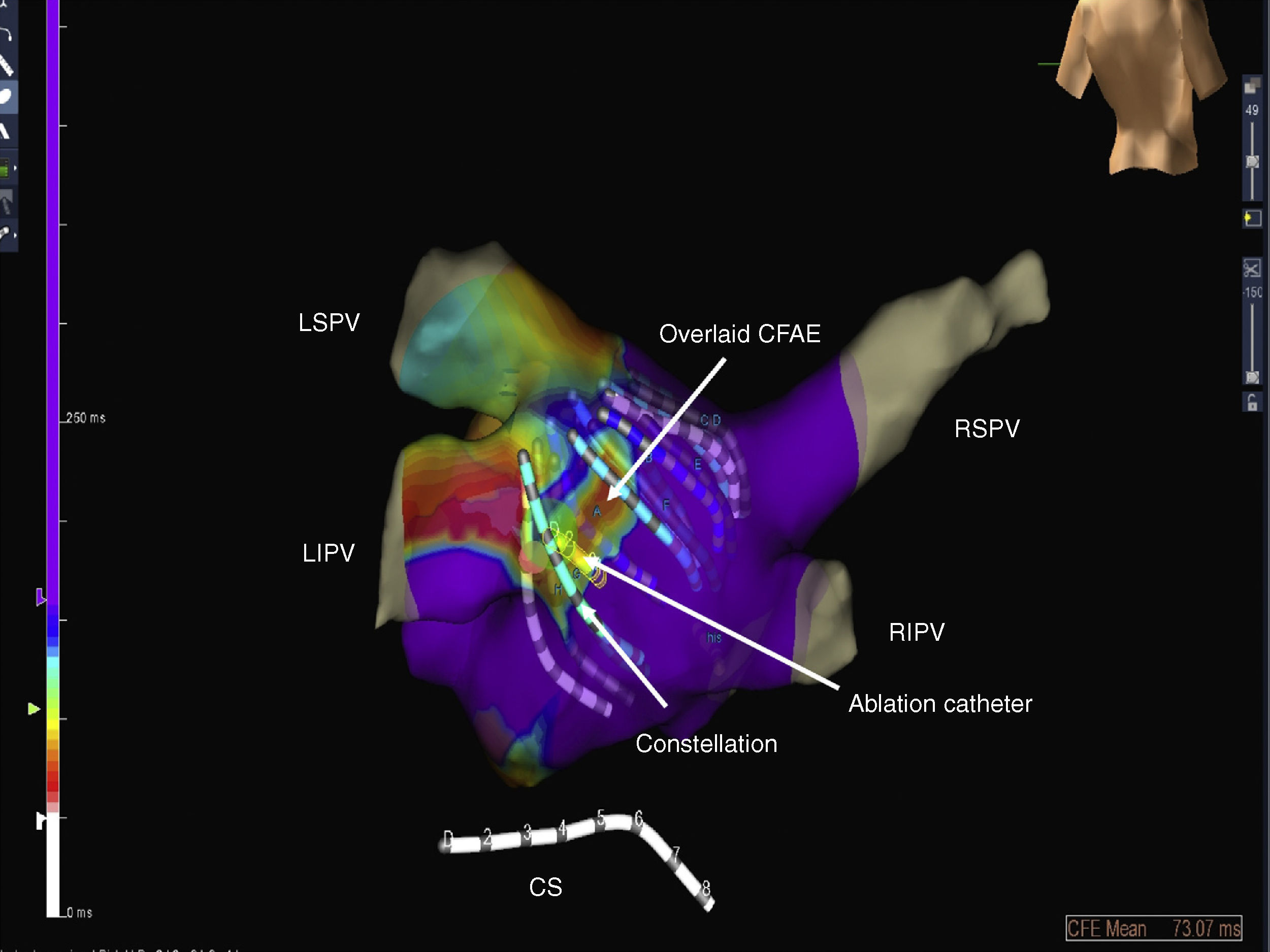
 defined by dynamic overlaid mapping. Anatomy defined by the Ensite NavX™ system; electrograms recorded by basket catheter (Constellation, Boston Scientific); ablation performed by ablation catheter (yellow) located over the area of a stable CFAE (white arrow). CFAE: complex fractionated atrial electrogram; CS: coronary sinus; LIPV: left inferior pulmonary vein; LSPV: left superior pulmonary vein; RIPV: right inferior pulmonary vein; RSPV: right superior pulmonary vein.")
Posteroanterior view of the left atrium showing a stable complex fragmented atrial electrogram (CFAE) defined by dynamic overlaid mapping. Anatomy defined by the Ensite NavX™ system; electrograms recorded by basket catheter (Constellation, Boston Scientific); ablation performed by ablation catheter (yellow) located over the area of a stable CFAE (white arrow). CFAE: complex fractionated atrial electrogram; CS: coronary sinus; LIPV: left inferior pulmonary vein; LSPV: left superior pulmonary vein; RIPV: right inferior pulmonary vein; RSPV: right superior pulmonary vein.
. LIPV: left inferior pulmonary vein; LSPV: left superior pulmonary vein; RF: radiofrequency; RIPV: right inferior pulmonary vein; RSPV: right superior pulmonary vein.")
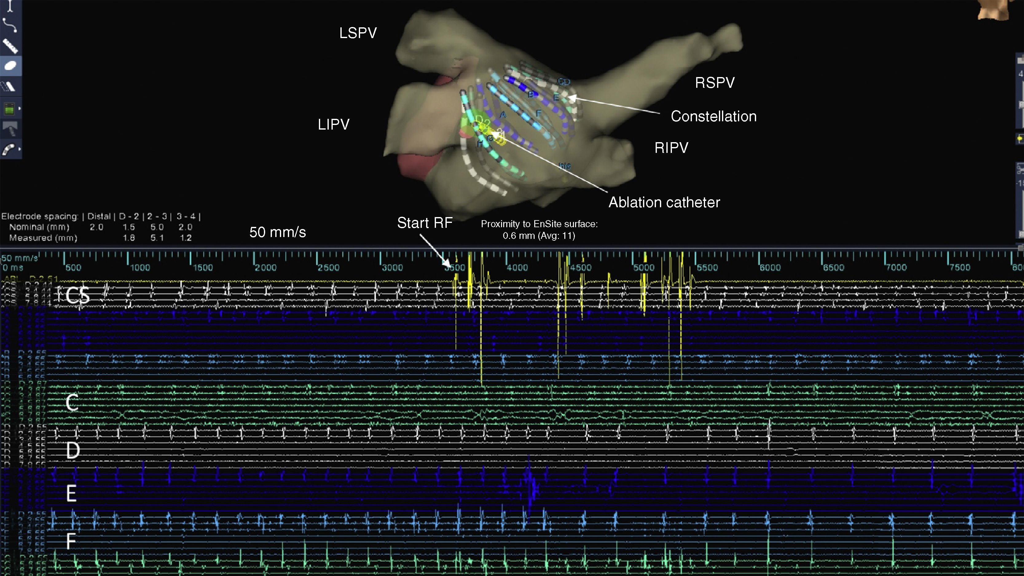
Above, anatomical mapping of the left atrium with the Constellation catheter and the ablation catheter. Below, coronary sinus electrograms and electrograms from the splines of the diagnostic catheter. The posterior wall is represented from splines C to F. Immediately after initiation of radiofrequency application, an increase in cycle length in the posterior wall is noted (scale 50 mm/s). LIPV: left inferior pulmonary vein; LSPV: left superior pulmonary vein; RF: radiofrequency; RIPV: right inferior pulmonary vein; RSPV: right superior pulmonary vein.

, interruption of the electrical activity of the posterior wall is seen (scale 25 mm/s to confirm continued electrical activity suppression). LIPV: left inferior pulmonary vein; LSPV: left superior pulmonary vein; RIPV: right inferior pulmonary vein; RSPV: right superior pulmonary vein.")
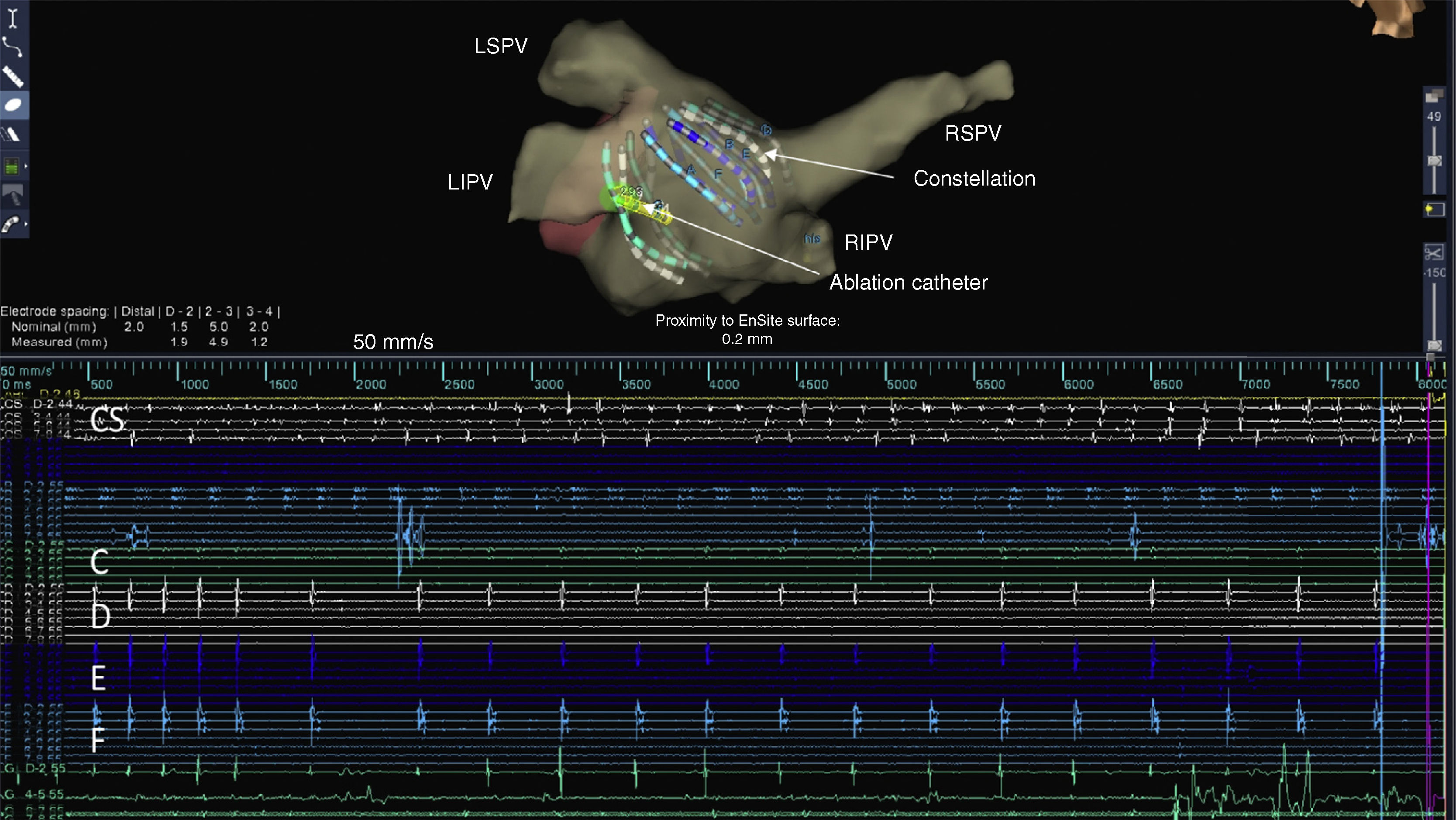
During the second radiofrequency application to eliminate the stable CFAE (enlarged lesion), interruption of the electrical activity of the posterior wall is seen (scale 25 mm/s to confirm continued electrical activity suppression). LIPV: left inferior pulmonary vein; LSPV: left superior pulmonary vein; RIPV: right inferior pulmonary vein; RSPV: right superior pulmonary vein.
The authors declare that the procedures followed were in accordance with the regulations of the relevant clinical research ethics committee and with those of the Code of Ethics of the World Medical Association (Declaration of Helsinki).
Confidentiality of dataThe authors declare that they have followed the protocols of their work center on the publication of patient data.
Right to privacy and informed consentThe authors have obtained the written informed consent of the patients or subjects mentioned in the article. The corresponding author is in possession of this document.
Conflicts of interestThe authors have no conflicts of interest to declare.
